Innovations In Trichology

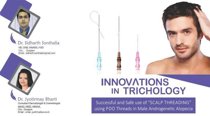
Successful and Safe use of “SCALP THREADING” using PDO Threads in Male Androgenetic Alopecia

Disclaimer: A part of this study has already been accepted for publication in the Journal of European Academy of Dermatology & Venereology, vide (MS no. – JEADV-2017-0505.R1)

Bharti S, Sonthalia S, Patil P, Dhurat R. Scalp threading with polydioxanone monofilament threads: A novel, effective and safe modality for hair restoration. J Eur Acad Dermatol Venereol [In Press]

INTRODUCTION

Androgenetic alopecia (AGA) results in significant disturbance in self-perception and psychosocial interaction in both genders. Although various treatment modalities including topical minoxidil (MNX), oral finasteride (FIN), synthetic bio-peptides, platelet rich plasma (PRP) and many others have been used with good therapeutic benefit, they either show suboptimal response or the response tends to plateau off with no further gain in hair regrowth.1 Hair transplantation is often not a acceptable to many patients owing to it being a surgical modality and/or the cost involved.

Polydioxanone (PDO) threads have emerged as a popular procedure for non-surgical face lift. Multiple modes of action have been suggested and confirmed histologically, including a foreign body reaction-induced neocollagenosis, mechano-transduction which is the fibroblastic response such as actin polymerization and matrix deformation in response to mechanical stimuli, and finally regulation of gene expression, cell differentiation, microcirculation stimulation, and promotion of tissue growth.2

Microneedling using a dermaroller, is another efficacious facial rejuvenation procedure that creates micropunctures leading to a wound healing cascade with release of various growth factors such as platelet derived growth factor (PGF), transforming growth factor alpha and beta (TGF-α and TGF-β), connective tissue activating protein, connective tissue growth factor, and fibroblast growth factor (FGF).3,4 This effect has been extrapolated to stimulate hair growth, confirmed by the results of a randomized double blind trial.4 Akin to the extension of microneedling from facial rejuvenation to stimulation of hair growth procedure, we attempted to assess the response to the insertion of PDO threads into the scalps of 5 male patients with Grade 3-4 AGA, and evaluated the role of this technique as a therapeutic modality for AGA.

PATIENT SELECTION

Based on the above premise, we evaluated the efficacy & safety of PDO thread insertion into the scalp in five male patients of AGA not responding satisfactorily even after 18 months of therapy with both MNX (10% lotion twice-a-day) and oral FIN (1 mg daily). A detailed patient counseling was done regarding the procedure, expectation of results, post-procedure care and expected time of any appreciable response (at least 6 months after the procedure), and written consent was taken. All patients were already off from both the treatments, i.e. oral FIN, and topical MNX, for at least 3 months prior to enrolment for the PDO threads insertion, and they were advised to maintain abstinence. The procedure was undertaken after ruling out diabetes and infections including HIV, Hepatitis B, hepatitis C, and syphilis. None of the patients had any significant systemic or metabolic disorder, keloidal tendency or on anti-platelet or any other drugs. In all patients, hair loss and recession were confirmed to be of AGA origin by detailed history, physical examination and trichoscopic analysis. The hair pull test was performed to rule out active effluvium.

EFFICACY EVALUATION

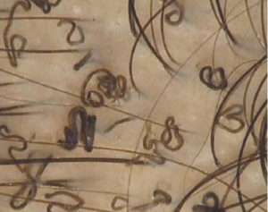
Global pictures of the frontal scalp, vertex, lateral and back view were taken before and after the procedure for comparison. After baseline global photographs, the scalp were shaved off to ensure equal length of hair shaft in all. Hair count was done trichoscopically in 1 cm2 targeted fixed area at baseline and at end of therapy (week 12). The primary end-point of improvement evaluation was the global photographic improvement (GPI), with secondary end-points being: 1) comparison of the trichoscopic hair count in the target area, and 2) degree of patient satisfaction on visual analogue score (VAS) ranging from 1-10.

METHODOLOGY: Thread Insertion

After signed consent, topical anaesthetic cream was applied over the treatment area 1 hour prior to the procedure, following which the treatment area was thoroughly cleansed with spirit and povidone-iodine. Monofilament PDO threads (30 mm long) were then inserted in the intrademal plane, extending from a pre-decided entry point covering the maximum possible length per needle. Multiple needles were inserted at 1 cm spacing (Fig. 1) in a radial orientation to cover the affected scalp (Fig. 2). The total number of needles inserted per scalp depended on the extent of bald area to be treated ranging from 20-40. During insertion, the scalp skin was stretched by the surgeon’s non-dominant hand to make it taut, and the needle was inserted through the desired point by the dominant hand, ensuring that the needle remains as parallel to the plane of the scalp skin as possible. After insertion, the needles were withdrawn leaving the PDO threads in situ.

The patients were recommended oral antibiotics for 5 days, and suggested to use a mild shampoo after 48-hours. They were also cautioned against applying any other topical over the scalp. Follow-up visits were done at 2 weeks, 6 weeks and 12 weeks after the procedure.

HA

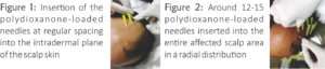
Figure 1: Insertion of the polydioxanone-loaded needles at regular spacing into the intradermal plane of the scalp skin.

Figure 2: Around 12-15 polydioxanone-loaded needles inserted into the entire affected scalp area in a radial distribution

RESULTS

Efficacy (Figs. 3 and 4)

There was no appreciable improvement till 6 weeks post-procedure. However, at 12 weeks, all patients (n=5) had variable but appreciable degree of increase in hair counts, with decent coverage of the previously bald areas. Global photography improvement (investigator evaluated) was significant in all patients resulting in 40-75 % with an average of 57%. The increment in the count of hair follicular units (HFU) by trichoscopy was observed in all patients. Hair count increased from average number of 48 HFU/cm2 to 93 HFU/cm2, with an average gain of 67 HFU per cm2. The patient satisfaction was good with VAS ranging from 4-8 with a mean of 6. The detailed results are mentioned in Table 1. Incidentally, the best outcome was observed in the youngest patient and the oldest patient in our series had the least improvement amongst the group.

Table 1. The results of scalp threading in 5 male patients with androgenetic alopecia at 12 weeks

HAHAHA

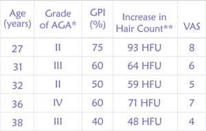
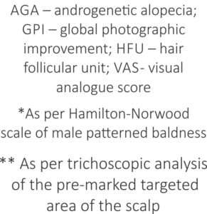
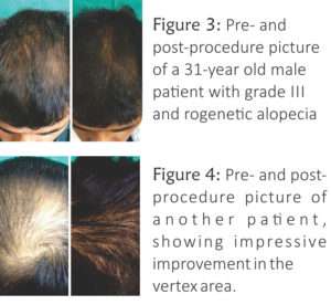
Figure 3: Pre- and post-procedure picture of a 31-year old male patient with grade III androgenetic alopecia

Figure 4: Pre- and post-procedure picture of another patient, showing impressive improvement in the vertex area.

Adverse effects

The procedure was very well tolerated by all the patients with the most common complaint being mild pain during thread insertion (n=5), followed by mild transient swelling (n=2) that resolved within 24 hours. There was no case of significant bleeding, ecchymosis, persistent pain, headache, or post-procedure infection. There were no other adverse effects till the last follow-up.

DISCUSSION

Polydioxanone (PDO) filament is a synthetic absorbable suture which is prepared from polyester, poly (p-dioxanone). It has high flexibility, high retention strength, non-allergenic and a slow absorption rate (6-8 months). It carries no risk of bacterial colonization or infection. When used for non-surgical face lift, threads stimulate neocollagenosis within 2-3 weeks with clinical results expected to last for 2-3 years.1 Although these threads are also available as barbed and screwed, we preferred monofilament threads.

The exact mechanism of action of hair growth stimulation by PDO threads remains speculative. However, it is likely that it is similar to that of microneedling, probably involving enhanced expression of hair-related genes, and release of growth factors like PDGF, and possibly, direct activation of stem cells in the hair bulge area.3,5  A recent randomized evaluator blinded trial by Dhurat et al. had indeed displayed the superiority of the combination of microneedling with MNX versus MNX alone in MPHL (grade III vertex or IV) patients.4

Although this pilot study seems to offer scalp threading as a novel efficacious and safe non-surgical approach to hair regrowth, further research with a properly designed controlled trial with a larger cohort is warranted to elucidate its role in the therapeutic armamentarium of hair restoration. The limitations of this study include the small number of cases, limited duration of follow-up period, and lack of histological analysis of the scalp skin post-thread insertion. Further, unlike the predictable time span of the effect of threads in facial rejuvenation, the duration of the hair growth stimulating effect of the threads remains to be explored. Also, one needs to be cautious about the development of foreign body granuloma in any PDO inserted patient.

References

  1. Sonthalia S, Daulatabad D, Tosti A. Hair Restoration in Androgenetic Alopecia: Looking Beyond Minoxidil, Finasteride and Hair Transplantation. J Cosmo Trichol 2016;2:1-13.
  2. Suh DH, Jang HW, Lee SJ, et al. Outcomes of polydioxanone knotless thread lifting for facial rejuvenation. Dermatol Surg 2015;41:720-5.
  3. Jeong K, Lee YJ, Kim JE, et al. Repeated microneedle stimulation induce the enhanced expression of hair-growth-related genes.Int J Trichology 2012;4:117.
  4. Dhurat R, Sukesh M, Avhad G, et al. A randomized evaluator blinded study of effect of microneedling in androgenetic alopecia: a pilot study. Int J Trichology 2013;5:6-11.
  5. Kim BJ, Lim YY, Kim HM, et al. Hair follicle regeneration in mice after wounding by microneedle roller.Int J Trichology 2012;4:117.

Hyaluronic Acid: An Underused Wonder Molecule

Hyaluronic acid (HA) is a very versatile molecule, with a ubiquitous presence in the human body.

It is found in the vitreous humour of the eye, the synovial fluid, extracellular matrix, bones, cartilage, umbilical cord, neural tissue and most importantly, the skin, where 50% of the total body HA is.

CHEMISTRY AND PHYSIOCHEMICAL PROPERTIES:

HA is a nonsulfated glycosaminoglycan, a polymer of disaccharides, present in the extracellular fluid. Polymers of hyaluronic acid can range from 5,000to 20,000,000 Da in vivo. It thus has a high molecular mass. It is present in all vertebrates, with the rooster comb containing the highest concentrations ever reported in animal tissues. It is also present in the capsules of microorganisms.

A 70kg adult has approximtely 15g of HA.

HA

PHYSIOLOGICAL FUNCTION IN THE SKIN:

The biological functions of hyaluron are related to the size of the molecule. Higher weight polymers cause immunosupperssion, filling (dermal fillers) and are antiangiogenic. Medium-sized polymers help in wound healing. The oligosaccharides are infammatory, immuno-stimulatory and angiogenic. The smallest polymers (400to4000Da) are antiapoptotic.

Hyaluronic acid is the major molecule that maintains hydration in the skin.

1g binds with 6L of water.

The human body has approximately 15g of hyaluronoc acid, one-third of which is “turned over” every day.

It is responsible for maintaining the turgor, elasticity and hydration of the skin.

It plays an important role in the synthesis of ECM molecules and in epidermal cell interaction.

It aids in wound healing and reduction of inflammation as mentioned above.

It modulates cellular immunity by preventing infections and reducing the chance of allergies.

It maintains the tissue homeostasis.

It acts as an immune regulator in human diseases.

It is highly biocompatible; and this property has made it a ubiquitously used wonder molecule.

SYNTHESIS OF HYALURONIC ACID:

In living organisms, it is synthesised by hyaluron synthtase enzymes.

HA

DEGRADATION OF HA

It has a dynamic turnover rate, with a half-life of less than a day in the skin. It is degraded by enzymes called hyaluronidases. It can also be degraded non-enzymatically by free radical mechanism, in the presence of ascorbic acid, ferrous or cuprous ions in the presence of oxygen.

Hyaluronic acid gets depleted with age.

Substances with polysaccharide-like structure have inhibiting effects on the degredation of HA by competing with it.Cross linking helps to prevent degredation to some extent.

It gets washed off with water and has to be re-applied.

HA and the SKIN

Hyaluronic acid is that wonder molecule, which keeps the skin firm, supple and hydrated. It maintains the moisture balance of the skin, and maintains the skin health.

It can adjust the moisture absorption rate based on the humidity in the environment. It thus maintains the skin health.

It is an important anti ageing molecule. It is used for fine lines on the skin.

Hyaluronic acid is said to have antioxidant and free radical scavinging activities.

It protects the skin from UVB rays.

HA is said to decrease the lipid concentration in the sebaceous glands.

HA AND WRINKLES

Hyaluronic Acid as an antiageing requires special mention. It combines with the keratin of the skin via hydrogen bridges in a water leaves a tension in the skin, which obliterates the fine wrinkles.

ENHANCING PENETRATION OF HA

Shorter chain polymers are better absorbed by the horny layer of the skin. Using an electroporation or ultrasound with itenhances the breakdown into shorter chains. Combining HA with a matrix of dextrin or modified cellulose too helps.

TYPES OF HYALURONIC ACID PRODUCTS

The biggest advantage of this molecule is that it can penetrate the upper skin layers and cross the skin barrier and it is an inert molecule.

The various forms available are

  • Serums
  • Creams
  • Oral supplements
  • Dermal cross-linked fillers (pre-filled syringes)
  • Auto cross-linked hydration syringes
  • As a powder for consumption
  • As an ingredient in mouth washes

THE FUTURE OF HA MOLECULE

The commercial value of hyaluronan is huge. It is sold for about 1,00,000 USD per Kg! The world market is around 500million USD. It is being used for diverse applications. It is already being used for cancer therapy. With newer insights into production techniques, and knowledge about is biological functions, this wonder molecule is here to stay, and being researched more and more to be put to therapeutic use. Nanoparticles and liposomes of hyaluronic acid are being developed to enhance its penetration.

Basics Of Dermatoscopy

Introduction:

The use of a dermatoscope in clinical practice has been shown to increase diagnostic accuracy and is considered the standard of care in assessing patients with pigmented skin lesions. Its use is also being increasingly applied to the diagnosis of nonpigmented skin lesions. Dermoscopy has been shown to facilitate the clinical recognition of several inflammatory and infectious diseases, as well as their discrimination from skin tumours.With new evidence continuously being gathered, the dermatoscope gradually acquires a role similar to the stethoscope of general practitioners, becoming an irreplaceable clinical tool for dermatologists Application of dermoscopy should follow the standard procedure of acquiring information from patient history and clinically evaluating the number, location and morphology of the lesion(s).

Four parameters should be assessed when applying dermoscopy in the realm of inflammatory and infectious diseases:

  1. morphological vascular patterns;
  2. arrangement of vascular structures;
  3. colours; and
  4. follicular abnormalities,

while the presence of other specific features (clues) should also be evaluated.

a

b

Figure: Difference between nonpolarized and polarized Dermatoscopy.

How do I interpret the dermatoscopic image?

There are only five basic elements used to describe pigmented structures, which are objectively defined using the following geometric terms (Figure 1):

  • line: a two dimensional continuous object with length greatly exceeding width
  • pseudopod: a line with a bulbous end
  • circle: a curved line equidistant from a central point
  • clod: any well circumscribed, solid object larger than a dot; clods may take any shape
  • dot: an object too small to have a discernable shape. Lines are further classified into five types: reticular, branched, parallel, radial and curved, as these have diagnostic significance.

c


Figure : Structures seen
with a dermatoscope lines (A), pseudopods (B), circles (C) clods (D), dots (E), Lines can be reticular (F), branched (G), parallel (H) radial (I), Curved (J)

Blood vessels are also described using similar well defined Terminology. (Figure).

d

Figure. Vessel patterns :
Vessel Structure (left): dots (A) clods (B), lines (C), looped (D), curved (E),serpentine
(F), helical (G), coiled (H)
Vessel arrangement (right): random
(A), clustered (B), serpiginous (C), linear (D),centred (E), radial (F) reticular (G), branched (H)

Colour has great diagnostic significance in dermatoscopy. The main pigments are melanin and haemoglobin, and the colours produced are shown in Figure 3. Although melanin is black, it appears as different colours depending on how deeply in the skin it is located.Very superficially it looks black, but deeper in the epidermis it appears brown. In the dermis, melanin appears either grey or blue. Therefore in many circumstances, these colours indicate the presence of melanin pigment deeper in the

skin than expected, which can be a clue to invasive malignancy.

e

Figure. The interpretation of colours seen with a dermatoscope

Features of Dermatoscopy in Inflammatory and non-inflammatory diseases.

Papulosquamous dermatoses:

Lichen planus;

Dermoscopy enables the visualization of Wickham striae

(WS), which have been assessed as a highly sensitive and specific criterion for the diagnosis of lichen planus (LP).Wickham striae are seen as round, linear, reticular or annular pearly-whitish structures and may develop thin or broadarboriform projections, surrounded by dotted or linear vessels that highlight them.

Psoriasis;

Dermoscopy of plaque psoriasis (PP) typically reveals white scales and regularly distributed, dotted vessels on a light red background ( 2b and 3a). Detection of other morphological types of vessel should raise doubts about the diagnosis of PP.

Under higher magnifications (100–400), the psoriatic vessels appear as convoluted loops, mirroring the underlying histopathological finding of spiralled capillaries within the dermal papillae, associated with the psoriasiform epidermal hyperplasia.10 Although present in every psoriatic plaque, red dots are not a specific dermoscopic feature, as they can also be found in other papulosquamous dermatoses. Instead, their uniform, symmetrical, regular distribution throughout the lesion typifies PP. When an intense hyperkeratosis impedes the visualization of underlying structures, scale removal will bring to light the aforementioned vascular pattern, possibly together with tiny red blood drops (the dermoscopic ‘Auspitz sign’). Other patterns of vessel distribution are extremely rare in PP, with the exception of the highly specific, but relatively uncommon, pattern of ‘red globular rings’.

Dermatitis

Dotted vessels in a patchy distribution and yellow serocrusts represent the main dermoscopic criteria in all subtypes of dermatitis (Figs ). Depending mainly on disease duration, dermatitis may dermoscopically display a spectrum of scaling and vascular changes, with acute exudative lesions predominantly exhibiting yellow scale crusts (‘yellow clod sign’), whereas more chronic and lichenified lesions are typified mainly by dotted vessels in a patchy distribution within the lesion.

Pityriasisrosea:

Both the herald patch and the secondary lesions of pityriasis roseadermoscopically display peripheral whitish scales (‘collarette sign’), typically combined with dotted vessels that lack the regular distribution of psoriasis.

f

Lichen planus (a) may share clinical features with other papulosquamous skin diseases, such as dermatitis (c). However, dermoscopic detection of Wickham striae (b, arrows) allows a safe diagnosis of lichen planus, while dermatitis (d) typically shows dotted vessels with a patchy distribution and yellow scale crusts (arrows).

Dermoscopic differentiation between psoriasis and dermatitis;

g

Dermoscopic differentiation between psoriasis (a, b) and dermatitis (c, d) is enabled by recognition of the white scales of the former in contrast to the yellow serocrusts of the latter.

Dermatoscopic criteria for inflammatory skin diseases.

h

Dermatoscopic findings of Infectious skin disease:

i

c

Dermatoscopic finding in Hair disorders:

Alopecia Areata: yellow dots, black dots, exclamation mark hairs, and vellus hairs.

k

Figure: Dermoscopic features of alopecia areata; yellow dots (A; arrow), clustered vellus hairs (B; dotted circle), black dots (C; triangle), and tapering hairs (D; white arrow).

Dermatoscopic finidngs in tineacapitis:

Comma hairs, cadaverized hairs, coiled hairs, haemorrhagic spots, honeycomb pigment network, yellow and black dots, thin hairs , corkscrew hairs, broken hairs, peripilar scaling etc.

i

Figure: Video demoscopy 40x.comma and corkscrew hair in tineacapitis.

Three point check list in pigmented lesion to detect early melanoma:

The dermoscopy, 3-point checklist for early detection of skin cancer is fairly easy to learn and has a high sensitivity for melanoma. There a high likelihood of malignancy (melanoma or basal cell carcinoma) if a pigmented skin lesion has any two of these criteria. The 3-point checklist has been designed to allow non-experts not to miss detection of melanomas.

  1. Asymmetry: asymmetry of colour and structure in one or two perpendicular axes
  2. Atypical network: pigment network with irregular holes and thick lines
  3. Blue-white structures: any type of blue and/or white colour, i.e. combination of blue-white veil and regression structures.

Dermatoscopes and dermatoscopy

A dermatoscope is more than a magnifying lens and light source. By eliminating reflection from the skin surface, the dermatoscope allows better visualisation of the patterns formed by pigment and blood vessels – critical features in the diagnosis of skin lesions. This can be done using fluid to couple the lens to the skin or by cross-polarisation of light source and lens. Colour rendition varies with the different types of instruments,9 hence familiarity with your chosen device is important.

Conclusion

In summary, dermoscopy improves the clinical recognition of several inflammatory and infectious dermatoses, by enhancing the most basic of diagnostic functions in dermatology: visual the dermatoscope gradually acquires a role similar to the stethoscope of general practitioners, becoming an irreplaceable

clinical tool for dermatologists inspection.


Cheyletiellosis ( Walking Dandruff ) : A Case Report in 2 Patients

Abstract:

Walking  dandruff  is  the common name for a skin condition caused by infestation of a dog’s skin by tiny Cheyletiella mites. These mites burrow through the outer layers of the dog’s skin, causing irritation and mild to severe itchiness as a result of mechanical irritation. Dogs, cats, horses and rabbits can become transiently infested with the Cheyletiella mites that normally populate other hosts. This happens through direct contact between an animal carrying the mites and a member of a different susceptible species (dogs, cats, rabbits or even people). For example, a human can become infested by Cheyletiella yasguri mites as a result of petting an infested dog, which is the normal host for that particular species of mite.

Case 1 : a male patient  34 years of age presented to us with complains of itching  off and on in  the groins for 1 month. He had taken self medication with topical creams without any improvement. On examination  few minute erthytematous papules with excoriations were noticed over the groins & inner thighs. There were no  annular lesions suggestive of fungal infection, the genitals were not involved and there was no history of nocturnal itching .He gave history of having pet dog  at home. Baseline investigations were within normal limits. On skin scrape for KOH examination revealed the large mite with four pair of legs and prominent accessory mouth parts which terminated in hooks. Diagnosis of Cheyletiella Yasguri  was made based on the miscroscopic appearance of the mite. Patient  was treated with tab Ivermectol 12 mg with anti pruritic soothing lotion and antihistamine.  On follow up 15 days later he showed satisfactory improvement in both itching and lesions.

Case 2: a male patient of 32 years old presented to us with asymptomatic , well defined alopecia patch on the scalp with scaling. His baseline investigations were within normal limits. KOH examination was done from the scalp to rule out tinea capitis which was negative . However we found  the mite  Cheyletiella yasguri in the same field. He was given necessary treatment for  alopecia and ivermectol shampoo for single hairwash.

01

Discussion

Walking dandruff can be described as a form of mild dermatitis that occurs due to the presence of a mite called Cheyletiellosis on the scalp. These mites, are more commonly found in animals like cats, dogs, rabbits and horses, but can also spread to humans. The motion of the visible mites give the condition its unusual name, as the dandruff seems to walk around the scalp.

Walking dandruff is highly contagious, as Cheyletiellosis mites easily spread from one host to the other. It can be contracted by prolonged personal contact with an infected animal. The entire life cycle of the mite on one host is around 21 days. Once the infected animal is treated, the possibility of transference to humans is eliminated. Because animals do not necessarily show symptoms of the mites the owners do not realise that they are infected. It is noticed as whitish, dandruff like flakes on the hair, which seems to be moving around. Other signs and symptoms of walking dandruff include severe itching and blisters on the scalp, red bumps on the arms, trunk and buttocks. These symptoms usually disappear within a time span of 3 weeks, as people are not the preferred hosts for the mites. However, humans can experience a lot of pain and discomfort because of the mites. therefore walking dandruff should be treated as soon as possible. A number of different have shown to be effective including high dose ivermectin, milbemycin oxime, moxidectin, selamectin, fiprofil and pyrethrin shampoos. Infestations in cats and dogs can be prevented by routine administration of topical antiflea applications on a monthly basis.

References:

  1. 1.Arther RG. 2009.Mites and lice :biology and control.VCNA Small Amin Pract. 39:1159-712.
  2. Paradis M, Villeneuve A (August 1988).” Efficacy of Ivermectin against Cheyletiella yasguri infestation in Dog”. Can.Vet.J.29(8):633-635.
  3. Bakkers, E.J.M; Fain, A. (1972). :Dermatitis in man and in a dog caused by the mite Cheyletiella yasguri Smiley”.British Journal of Dermatology, 87(3):245-247.
  4. Powell, Ralph F;Palmer, Susan M;Palmer, Charles H; Smith, Edgar B. (1977). “Cheyletiella dermatitis”, International Journal of Dermatology.16(8):679-682.

Jaw Sculpting and the Neck rejuvenation

Since the beginning of time, a beautiful neck and well defined jaw are highly sought after features that signify youth, health, femininity and beauty. In the 14th Century BC, Queen Nefertiti was known for her striking beauty and sculpted neck lines. Today, women are frequently requesting a refined jaw line similar to Angelina Jolie or Jennifer Lopez. With the modern advancements in aesthetic medicine, there are many ways to achieve a more sculpted jaw line and smooth feminine neck without going under the knife.

Ageing and anatomy: The aesthetic anatomy of the neck can be divided into several layers, from superficial to deep, starting with the skin, subcutaneous tissue, superficial muscular-facial layer and deep subplatysmal structures. Senescent changes of the mature neck include accumulation of fat, laxity of muscular support, and the cumulative effects of photodamage and gravity. These contribute to the loss of definition of the cervicomental angle, submental fullness, sagging of the jowls, inelasticity and redundancy of the skin, along with platysmal band formation. As a result of these developments, the lower facial third may appear fuller, which can diminish the ‘heart-shaped’ facial shape that is so widely associated with a youthful appearance. The minimally invasive, nonexcisional interventions includes laser, light, radio frequency, high-intensity focused ultrasound (HIFU) energy-based therapy ,injectable soft tissue fillers, neuromodulators, and ablative and nonablative technologies for skin rejuvenation, as well as suture-based suspensory techniques, all used alone or in combinations can help in rejuvenation of this area.

Important limitations to treatment include the anterior location of the thyroid and parathyroid glands, which must be shielded from deeply penetrating wavelengths. There is increased scarring risk on neck and chest as compared to face, thus necessitating greater care and lower fluences in this region.

Understanding a patient’s concern and cosmetic goals is key to successful treatment. The treating physician should be aware of their general health, diet and lifestyle factors, medications used, history of treatments, skin care routines etc. The patients should be asked the questions on how much the problem bothers them and whether or not they would consider cosmetic surgery, which can help to establish what type of treatments to offer the patient. All non-surgical skin tightening and rejuvenation treatments require the patient to be able to commit to a consistent skincare regime in order to enhance and maintain results. Patients should know why they should wear a daily SPF and why their smoking habit might be impending their results. Once we have the basic education in place the rest of the dialogue is easy. Next, a decision should be made on what treatment to offer, this is often dependent on the patient’s budget. Injectable treatments in combination with other procedures can help patients achieve dramatic non-surgical results.

Baseline and follow-up photography from both sides is exceedingly important, as the degree of improvement in neck laxity is often best appreciated by side views. It is recommended that the baseline and first set of follow-up photographs be reviewed with the patient if the level of efficacy is in question. Patients are cautioned that in the vast majority of cases, multiple treatment sessions are required to achieve significant tightening, depending on the technology used.

Nefertiti Lift: Jaw sculpting and neck lift using injectables are described in consumer media often as the ‘Nefertiti Lift’. For many people, the combination of injectable filler and botox can be a perfect nonsurgical solution to achieve a refined, elegant and defined neck and jawline.

Injectable fillers work extremely well for enhancing the jaw line. By adding volume, we can build upon the existing bone structure and create a stronger, more defined line. The injections are placed along the jaw bone in the areas where enhancement is desired. Side effects may include mild bruising and swelling for a few days. It is recommended to avoid anything that has a blood thinning effect on the body for at least one week prior to injections. It is also helpful to take arnica and/or bromelain tablets for a few days prior to treatment as well as a few days following (or as directed).

After the filler is injected into the desired areas, we can further improve overall appearance, sculpting the jaw and neckline with botulinum toxin injections. This procedure is done by injecting botulinum toxin around the jawline and down the neck into the platysma muscle, which is responsible for the downward pull along the jaw. By relaxing this muscle, the muscles on the upper face become stronger and naturally lift the face upwards. The end result is a youthful, tighter, more contoured appearance. These injections are more delicate than upper face botulinum toxin treatments and require an experienced operator who has excellent knowledge of the muscles of these regions, their functions, the antagonist actions exercised on other muscles, particularly in terms of the complex equilibrium of the mouth. An excessive dose, an inappropriate injection point, or a centering mistake can all easily be responsible for undesirable side effects. However, the results obtained, often with lower doses than in the superior part of the face, can be highly satisfactory.  A series of 3 to 5 botulinum toxin  injections are placed  1-2 cm apart on a horizontal line under the mandible posterior to the hypothetical line were the nasolabial fold meets the mandible. If present, injection of each platysmal band every 2 cm with 2 to 4 injection points per band (injection of platysmal band will be done by holding the band between 2 fingers and injecting intramuscularly, doing so ensures that you are only injecting the platysmal band; you don’t want to inject the deeper muscles in the neck as they could affect swallowing) A total of 20 to 30 units of botulinum toxin type A is used. Follow up will be done at 15 days for retouching or for post op pictures if no retouching is needed. If retouching is needed post injection pictures will be taken 10 days post retouching. Botulinum toxin can be injected every 4 to 6 months in order to maintain the results. It is not recommended to repeat the injections any sooner because of the risk for antibody formation and muscle-atrophy. Very dilute hyaluronic acid gels in the subdermal space, as well as PRP (patelet rich plasma) and other stem cell injections, have also reported to provide reasonable rejuvenation of the neck.

Chromaphore-based pathologies of the neck is common, given its sun-exposed location on the head and neck region. Topical therapies including bleaching agents, peeling agents, sunscreens , chemical peels, mesotherapy, chromophore-based lasers and light-based sources (532-nm wavelength Potassium titanyl phosphate (KTP) lasers, 694.5 nm Q-switched Ruby, and the 755 long-pulsed or Q-switched Alexandrite lasers, Pulsed dye lasers in the 585-nm wavelength and IPL ) can be used in different combinations. Generally, for neck rejuvenation in skin types I, II, and III, with dyschromia, cutoff filters in the 515-nm to 580-nm range have been very successful. For skin types 4 and 5, long wavelength cutoff filters in the 590-nm to 640-nm ranges, lower energies, and longer pulse configurations have allowed the treatment of darker discoloration in patients with more advanced  Fitzpatrick skin type. It is important that the IPL settings are gentle moderate in fluence, as IPL may induce a permanent hypopigmentation or discoloration of the skin.  Fractional nonablative and ablative lasers, and ablative fractional radiofrequency devices has also provided an opportunity to improve dyschromia and photoaging, as well as fine lines and texture of the neck.

Complications of the management of melanin and dyschromia of the neck include scars from overzealous laser and light-based settings, hypopigmentation from aggressive settings that result in a complete or near-complete clearance of melanocytes, as well as demarcation from treated and untreated areas.

2

Vascular or hemoglobin-based neck rejuvenation: The vascular proliferation derived from photoaging responds very nicely to the intense pulsed light, deep dermal and subdermal, proliferative vascular lesions occur in the neck and monochromatic long-pulse or variable pulsed wavelengths, such as long-pulsed neodymiumYAG or short-pulse and long-pulse, pulsed dye lasers are required.

Procedures such as simple shave excision, chemical or thermal ablation of intra-epidermal papillomas, skin tags, compound moles, seborrheic keratosis, actinic keratosis, and a host of other pathologies can significantly improve the appearance of the neck. Superficial and deep dermal epidermal rhytides can be treated safely and effectively with fractional ablative lasers, CO2, Erbium, and fractional radiofrequency ablative systems. 

Dermal and Subdermal Tightening Devices and Technologies: There has been a rapid evolution in our ability to provide moderate, non surgical skin tightening and wrinkle-reduction therapy with transepidermal energy devices. These new “energy-assisted” nonsurgical skin tightening procedures have become very important drivers of consumer interest. The first generation of the noninvasive skin tightening technologies involved nonfractionated longer wavelength near infra-red laser devices, such as the 1320-nm Cooltouch (Roseville, CA), the 1440-nm Smoothbeam (Syneron Candela, Yokneam, Israel), the long-pulsed Nd:YAG, and the1320 to 1440 nm synchronously pulsed Affirm MPX (Cynosure, Westford, MA).  Monopolar, stamping RF  typified by Thermage (Solta Medical, Hayward, CA), was a next generation very successful device with modest to good skin tightening effects, proven in large multicentered trials. Monopolar thermage protocols for treatment of the neck often includes 2 to 3 passes and 2 to 3 treatment sessions separated by several weeks. Combined optical-bipolar RF devices emerged, such as the Refirm and Polaris (Syneron), showed noticeable improvements using multiple-pass, multiple-session treatment protocols. These mono-polar and bipolar RF or optical-RF combination devices, are “stamping” or “static” in nature and often suffer from inadequate dermal stimulation by a combination of very high peak dermal energy (and hence stimulation) but a very short pulse duration, exposing dermal tissue to a relatively short thermal stimulation that would be required for the production of new collagen, elastin, and ground substances. These stamping devices generally deploy protocols with multiple passes and multiple treatments to overcome the ultrashort pulse duration but high temperature model of collagen production stimulation.  More recently, a whole class of transepidermal RF heating devices have emerged that are not short-pulse duration “static” or stamping in nature, but rather, are continuous wave RF systems that are constantly moved along the surface of the skin along a thin layer of ultrasound or some interface gel. The advantage of these “moving” or “dynamic” RF systems is the ability to heat this tissue to a lower temperature but for a much longer period than pulsed mode stamping technologies and, depending on the “moving” device, the therapeutic thermal end point, usually 42ºC to 43ºC can be maintained, for a very long time. Some of the early “moving RF systems” include the Accent (Alma lasers, Buffalo Grove, IL), Tripolar (Polagen), the diamond polar and Octapolar (Venus Freeze [Venus Concept, Toronto, Canada]), the Excelis (BLT Industries Inc, Framingham, MA), and  moving bipolar thermally controlled and modulated RF device, called the FORMA (Invasix). The FORMA is a very high tech, thermally modulated enhanced moving RF heating device that has built within the hand piece sensors that measure high and low dermal impedance, epidermal temperature, and electrode contact 10 times every millisecond, and automatically adjusts RF energy depending on the sensory feedback. The FORMA will automatically cut the RF energy off when the therapeutic skin temperature is reached, the impedance drops too quickly (temperature is rising too quickly), or the electrodes lose contact with the epidermal surface. Once the epidermis cools to 0.1ºC below the target temperature, the RF energy is turned on again and heating resumes. It can read, modulate, and automate the high and low temperature extremes, keeping the skin at a very uniform and consistent thermal end point, usually 42ºCto 43ºC for prolonged periods of time by this process of thermal modulation and eliminating the “hot spots” that can cause patient discomfort and burns. This thermomodulation process is called ACE, or acquire, control, and extend. Clinical and histological studies using ACE RF devices have shown good contraction and 14% more new collagen, and 35% collagen synthesis up-regulation. Infrared Light (1,100–1,800 nm, Titan, Cutera) have also shown good skin tightening results. Over the past few years, fractional deep dermal ablative devices have been released and commercialized that can result in significant rejuvenation. Ulthera, or fractional HIFU, uses high-frequency focused ultrasound to create ultrasound-induced fractional thermal ablative zones in the deep dermis and, in some areas, the superficial aponeurotic system. Results can be excellent, but occasionally painful and inconsistent. The HIFU can be combined with IPL or other fractional ablative devices at the same session. Deep RF ablative needle devices are also commercially available, which uses RF-emitting needles inserted under local anesthesia to create deep microthermal ablative RF zones that result in remodeling and tightening, while sparing the epidermis, hence risk of postinflammatory hyperpigmentation. The skin-tightening results of the these fractionated, vertical HIFU, or RF systems can be excellent to good, with, in general, one maintenance treatment every 3 to 6 months. Thermally modulated nonablative skin-tightening applicators also can be used safely off the face and in combination with any other injectables and chromophore based laser systems of fractional, ablative RF or laser systems. 

Excessive preplatysmal fat will often compromise a youthful, acute cervicomental angle. There are multiple ways to address submental adiposity in a minimally invasive fashion likes suction-assisted lipoplasty (SAL), ultrasound-assisted lipoplasty (UAL),  laser-assisted lipolysis (LAL) , injection lipolysis using drugs generally based on phosphatidylcholine and deoxycholate (PCDC), carboxytherapy etc.

The aging neck remains one of the greatest challenges for the aesthetic physician. Minimally invasive, nonexcisional techniques to rejuvenate the midface and brow have delivered tremendous success for  over the past 5 to 10 years. Because of its structure, location, and, often, sun exposure, the cervical submental region has presented more challenges to the aesthetic physician in achieving consistent nonexcisional rejuvenation. Over the past few years, with the evolution of subdermal heating techniques and transepidermal fractional ablative techniques, chromophore-based and light-based systems, alone on in combination with subdermal stimulation and suspension techniques, the aesthetic physician now has many weapons and tools to better address the noninvasive and minimally invasive, nonexcisional treatments of the aging neck.

As with any aesthetic treatment, appropriate patient selection and the skill of the physician are key to successful results. As with any machine, it’s only as good as the user. You have to choose the right patients and whoever’s using the machine has to be competent. Research your choice of machine carefully – there is a variety to choose from on the market and there’s one to suit every clinic according to patient requests and budget.  

In my practice I always use a combination approach for lower face and neck rejuvenation for my patients;  botulinum toxin and filler injections with few sessions of fractional radiofrequency and laser treatments,  infrared treatments and injection lipolysis using deoxycholate  and carboxytherapy when needed.

References

  1. Scarborough et al, ‘Exploring Aesthetic Interventions: Treating the Sagging Jawline and Platysmal Banding: A Simplified Technique’,The Dermatologist, 15 1 (2007)
  2. The ‘Nefertiti lift’: a new technique for specific re-contouring of the jawline. J Cosmet Laser Ther.2007 Dec;9(4):249-52
  3. Ultherapy, Harness the power of sound to non-invasively list skin, (US: Ultherapy, 2016)
  4. Neck rejuvenation Chapter 2 : Treatment of Neck Laxity with Radiofrequency and Infrared Light, Macrene Alexiades-Armenakas.
  5. Mulholland RS. An in-depth examination of radiofrequency assisted liposuction (RFAL). J of Cosmetic Surg and Medicine 2009;4:14–8.
  6. Duncan DI. The evolution of mesotherapy. Presented at the ASPS Breast and Body Contouring Symposium. Santa Fe (NM), Oct 4–6, 2012.
  7. Duncan DI. Injection lipolysis update. Presented at IMCA Asia. Hong Kong, Oct 4–6, 2012.
  8. Alam M, White LE, Martin N, et al. Ultrasound tightening of facial and neck skin; a rater-blinded prospective cohort study. J Am Acad Dermatol 2010; 62(2):262–9.
  9. Gotkin RH, Sarnoff DS, Cannarozzo G, et al. Ablative skin resurfacing with a novel microablative CO2. J Drugs Dermatol 2009;8(2):138–44.
  10. Taub AF, Tucker RD, Palange A. Facial tightening with an advanced 4-MHz monopolar radiofrequency device. J Drugs Dermatol 2012;11(11): 1288–94.
  11. Stampar M. The pelleve procedure: an effective method of facial wrinkle reduction and skin tightening. Facial Plast Surg Clin North Am 2011; 19(2):335–45.
  12. Hruza G, Taub AF, Collier LS, et al. Skin rejuvenation and wrinkle reduction using a fractional radiofrequency system. J Drugs Dermatol 2009;8(3): 259–65.
  13. Sadick NS. Update on non-ablative light therapy for rejuvenation: a review. Lasers Surg Med 2003; 32:120–8.
  14. Sadick HS, Sorhaindo L. The radiofrequency frontier: a review of radiofrequency and combined radio-frequency pulsed-light technology in aesthetic medicine. Facial Plast Surg 2005;21: 131–8.
  15. Mulholland RS. Radiofrequency energy for noninvasive and minimally invasive skin tightening. Clin Plast Surg 2011;38(3):437–48.
  16. Mulholland RS. Nonexcisional, Minimally Invasive Rejuvenation of the Neck.

Chronic Fungal Infection

A Female patient aged 55 yrs presented with large, erythematous, scaly symmetrical patches on both cheeks, few lesions on right side neck.

  • More than 3 yrs duration (on & off)
  • Associated with severe itching & burning sensation
  • Unable to cook food.
  • Not a diabetic (or) Hypertensive

Known case of vitiligo vulgaris > 20 ys duration took treatment long back, with less response.  Stopped medicines 5yrs back for vitiligo later she developed this rash for which she was roaming here & there with irregular treatment.

Initially it was treated as sun burn with sunscreens & steriod creams. the rash subsided to recur again.  Patient continued same medicine (steroid abuse) after 6 months she consulted another dermatologist.  Though treatment was given for fungal infection, patient had no symptom relief.  Irritation increased & discarded the medicine.

In April 2016, she presented to our clinic with symmetrical patches.  She had severe itching & redness (unable to come out from the house.)  Patient was vexed up with medicines and refused routine lab test also clinical diagnosis of Tineafacie with polymorphic light eruption (PLE) was made.  But counselling the patent was difficult her compliance to the treatment was doubtful.  We encouraged her for minimum 3 months regular treatment.

Treatment :

1st visit – oral terbinafine 250mg 21 days

  • 2% clotrimazole cream
  • anti histamines

2nd visit – rash got exacerbated

  • moisturisers 3 times daily
  • 2% clotrimazole cream night
  • oral itraconazole bd for 2 weeks
  • anti histamines orally

Counselling her was tough

3rd visit – Good improvement was noticed

  • 100mg itraconazole Bd
  • anti histamines
  • moisturisers topically
  • 2% clotrimazole cream night daily

Still under follow up recently her husband got tineafaciei.

We are all facing similar problems recurrent/persistant fungal infection

What can we do :

  • proper drug dosages orally & topically
  • avoidance of steroid added fungal creams
  • counselling the patients about the disease, proper storage of old slips
  • importance of continuing medicines for 2-3 months
  • monthly visits made compulsory

Though fungal infection are diagnosed easily, super added dermatoses must be identified and treated simultaneously.  In our case tinea + sun allergy were there couldn’t tolerate sunscreen hence simple moisturisers were added.  To see the results (symptom free) it took more than 2 months.

Adding moisturisers to the treatment made big difference.