How to be a Successful Medical Entrepreneur

When rising concerns over the country’s overburdened public healthcare system meet the growing emphasis on the role of entrepreneurship in achieving a self-sustaining and developed economy, it is medical entrepreneurship which guarantees the best of both worlds.

A large number of people have forayed into medical entrepreneurship with twin intentions of both earning profits as well as giving back to the society. This profession has gained tremendous popularity in India today.

Why most dermatologists are not great entrepreneur, how to act on this,


Today, thousands of business ventures are launched every year. Many never get off ground. Some grow at a slow pace, while others fizzle out after spectacular stat. Most of them start with a lot of enthusiasm and speed. But after a period of time, the organization stops growing.

Growth is not a monopoly of corporate only. Any dermatologist can and should visualize big picture. Goal should be bigger than life.

Generally, it is perceived that any doctor including Dermatologist cannot become good entrepreneur and second belief is entrepreneurship is to make more money what he is currently making.

However, this is just misconception as an individual Dermatologist becomes an entrepreneur to live an independent life where he no longer remains answerable to anybody. He is free to explore his creativity, work on his terms, create an empire of his dreams and leave a legacy in this world which will keep him eternally alive in the hearts of thousands of people and the independence to do all this comes through certain fundamental rights.

sakhiya sir

Every entrepreneur Dermatologist has certain rights that he/she possesses since the beginning of entrepreneurial journey till the time he leaves this world and these are known as the TEN ENTREPRENEURIAL RIGHT.

1) RIGHT TO DETRESS- Right to avoid all activities, relationships and circumstances that cause stress, anxiety and hostility and move from a world of distress to De-stress.

2) RIGHT TO MY TIME-Right to plan your time for yourself in a manner that is more productive and rejuvenating.

3) RIGHT TO WEALTH- Right to acquire the riches of the world for oneself, enterprise, associated people and entire echo-system by doing what you love to do and do best.

4) RIGHT TO FOCUS- Right to remain focused towards our most important goals, commitments, relationships and priorities amidst all the daily complexities taking place in our surroundings.

5) RIGHT TO STRATEGIES- Right to thoughtfully plan, schedule, execute activities and channelize all resources in a manner that you experience boundless growth

6) RIGHT TO ATTRACT- Right to attract the most lucrative relationships, resources, situations and alliances that will enhance the present status and lead to better future.

7) RIGHT TO THRIVE- Right to enjoy increased amounts of success, money and accomplishments without any guilt of having high aspirations and thereby thrive as a entrepreneur.

8) RIGHT FOR CAUSE- Right to go beyond yourself; live for a cause bigger than life and becomes the person you idealize.

9) RIGHT TO A BETTER TOMORROW-Right to visualize and work towards a better tomorrow

10) RIGHT FROM COMMODITIZATION -Right from commoditization by eliminating price war and yet winning business by creating value for every individual in each and every physical and intellectual intervention.

Like any other entrepreneur, a medical entrepreneur faces many problems in establishing the brand– be it financial and investment hurdles, rampant bureaucracy, abiding by corporate and government regulations, competition in the form of giant multinational corporations, and other challenges.

There are certain factors to be kept in mind in order to maintain a successful foothold in medical entrepreneurship: 

1.Assessment of needs through market research:

Before venturing out, medical entrepreneurs need to first understand what the market needs in order to tap into them. Healthcare is a vast field and it is through extensive and continuous research that medical entrepreneurs can choose the right Market demography. 

The local demand for a variety of health products and services have to be analyzed continuously so that entrepreneurs can carve a niche in an untapped market or even maintain good, profitable standards in a competitive market. 

To be successful, medical entrepreneurs need to find out where the gap lies in health services and then work to fill in those gaps, thus connecting with patients better.

For example, establishing yet another Dermatology Clinic in a city that already has a number of well reputed clinics/hospitals might not be too good an idea. A better way would be to look for regions that lack quality medical care and offer them quality healthcare solutions. This will not make the venture just financially successful, but would also make it more socially relevant. 

2. Choosing the right investors:

The right investor can make a big difference to your business. In the healthcare sector, it is an added bonus to find an investor with firsthand knowledge of that particular healthcare field. Even if medical proficiency is not guaranteed, someone with an insight into the current health economy is required for guaranteed long term growth and survival of the business.

Moreover, a big-name investor can help give you clinical validation and promote your brand. It is also very important to find someone who shares your vision, both for profitability and advancement in medical science. A friction with the investor in terms of goals and values might lead to major challenges in the long run. 

You can attract good investors in your field through adequate networking, association with medical and academic organizations and being well-equipped with market research and knowledge about the industry norms and opportunities. 

3. Balancing social responsibility and profitability:

One of the major moral dilemmas faced by the medical entrepreneur is the need to establish a profitable business while maintaining their social responsibility to general public health.

One must remember that there is a huge dearth of affordable health treatment and services in India, especially for the poor. There is also a widerural-urban gap in availability of medical professionals.

While medical entrepreneurs may not be able to break even if they take on the burden of free clinics for this purpose, an association with NGOs and regular voluntary work by employees in rural camps helps in fulfilling their social responsibility. 

For those looking to provide relatively affordable service in the private sector, cost effectiveness is integral to ensure that you make profit. 

Investment in medical research in order to assist the government with preventive and curative measures in significant health issues is also an important move to establish one’s social entrepreneurship. 

4. Constant technological innovation:

Innovation is the key to any successful entrepreneurial venture. Medical entrepreneurs need to keep up with the changing technological environment in healthcare and stay ahead with trends.

From electrical health records, telemedicine and digital marketing campaigns to advancement in medical technology which enhance patient treatments, health equipment, and surgical procedure, technological expertise is everywhere. 

One can uncover dormant or unmet needs by asking patients simple questions on how to improve their services. This way, one can gain competitive advantage through creative innovation. 

5. Knowing your limitations:

Don’t be tempted to take up a profitable line of service without having adequate skill and knowledge in that field. 

Keep in mind the ability of your team of medical consultants and know your limitations. Moving towards a specialization without the right human resources and training will be detrimental to your success. 

Patient’s feedback is very important as it will help highlight areas to improve upon. 

6. Playing to your strengths:

Ambition is what makes a successful entrepreneur. Every medical entrepreneur must set certain goals for themselves and the health industry and work towards achieving them by playing to their strengths. These could range from research and development in chronic diseases to enhanced customer service to a digital revolution in medicine.

In the end, remember that a bit of healthy competition is always good for the nation, especially when it results in saving lives.


Treatment Of Alopecia Areata Of Eyebrow With Non Needle Mesotherapy

ABSTRACT:

Alopecia areata of eyebrows are very resistant to routine treatments and takes long time for hair growth and the growth will be sparse and scattered and not in uniform length. Till the growth of new hair the patient will be in a state of anxiety and depressed and try to conceal the face and if necessary he will try to conceal and camouflage with hair dye and eyebrow pencil.

Mr. X aged around 40 years came with patchy hair loss area in left eyebrow of 6 months duration .he tried all native medicine including onion, but with no hair growth, the patient was assured and given following meso treatment. He was advised to apply triamcilone cream two time daily, along with mesotherapy with triamcilone cream alone 5 minutes in a week for about 12 week, after which he was advised to continue the triamcilone cream alone for another 12 weeks, he had been advised to come for review after another 12 weeks.

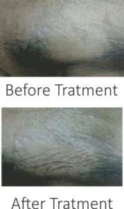
After 24 weeks patient came for review and found minimal hair growth with white hairs, which were dyed by the patient. Photos taken before and after treatment showed improvements in hair growth. The case report is given here for the following reasons.

  1. Hair growth in alopecia areata in eyebrows is very slow when compared
  2. Hair growth is accelerated and hastened by the mesotherapy of steroid cream without needles.
  3. This procedure is safe, painless and without side effects like secondary infections.
  4. This procedure speeds up the hair growth along with routine treatment.

alopecia

Cosmeceuticals & Dermatologists

Over last two decades, the cosmeceutical market has grown by leaps and bound. Not only is the number of cosmeceutical manufacturers and products on rise but the amount of money spent on skin care has also landed upward significantly.

As the science and industry of cosmeceutical grows, dermatologists now find themselves enjoying more opportunities to expand their treatment horizon and it also adds to the responsibility of being equally reasonable and justifiable while prescribing.

But aside from how they affect our business, cosmeceuticals are arguably changing the treatment landscape of our field. Skin care agents are more targeted and tolerable than before.

The term “cosmeceuticals” refers to the agents that facilitate skin care and while skin care is important for overall skin health, it is not a determining factor in managing any one dermatological condition.

Perhaps the most prominent example of this change is the case of the various topical forms of vitamin A, which has been used for more than 40 years in dermatology. It is the primary ingredient in topical tretinoin formulation for acne and photo – damaged skin for several decades. However, in recent years, we have seen a movement towards non-prescription retinols. Now, retional is a main ingredient in a variety of OTC products and has been combined with sunblocks and other bioactive ingredients like antioxidants to boost its clinical efficiency.

Many reputed pharmaceutical companies are coming up with products rich is nutrients and other such ingredients that go beyond basic skin care. They encompass ingredients like ceramides that mimic the skin barrier and are useful as repair agents. As such, these agents can potentially play a supplementary role in sun protection, and inflammatory conditions such as acne, eczema, and even psoriasis. These agents are not likely to displace topical steroid prescription agents but rather play a complementary role in treatment – nevertheless these products are actively blurring the lines between what was once a very clear divide between prescription agents and OTC products.

With all the advances in cosmeceutical technology, however they have a long way to go before being considered truly viable addition to a regimen.

The right cosmeceutical can be a good addition to a therapeutic or daily skin care regimen, but one of the most important aspects of integrating cosmeceuticals is keeping it simple. It’s our job to simplify regimen as much as possible. Given the lack of regulation and FDA guidelines for cosmeceuticals, manufacturer can essentially say anything about their products.

Many product lines have appealing packaging and a lot of media hype but fall short on actual benefits. It’s good to inform patient about the cosmeceutical market and how it differs from drugs. It makes them more discerning consumers. And also illustrates to the patient why your guidance is particularly valuable.

As skin care experts, we should be on the forefront of skin care revolution guiding patients on the selection and use of cosmeceutical agents may necessitate spending on extra few minutes with them, but it represents an excellent avenue to meaningfully influence their health and making a mark as a dermatology expert.

KHELLIN-An Old Wine In New Bottle

HISTORY

Khellin has been used as an herbal folk medicine, with use in the Mediterranean dating back to ancient Egypt, to treat a variety of maladies including: renal colic, kidney stones, coronary disease, bronchial asthma, vitiligo and psoriasis.

COMPOSITION

It is a major constituent of the plant Ammi Visnaga, also known as Bishop’s weed. Once purified, khellin exists as colorless, odorless, bitter-tasting needle-shaped crystals and is classified as a gamma-pyrone, a furanochromone derivative.

Chemical Structure

Khellin as a photosensitizer, a furanochromone extracted of the plant Amnivisnaga (5, 8 dimetoxi.2 methyl-4, 5 furo-6,7 chromone), and UVA irradiation (KUVA). The main advantage is its lack of phototoxicity, making it safe for use either as a home treatment or a treatment involving natural sunlight, even on a daily regimen.1 It is also less mutagenic than psoralens, and lessens the darkening of normal skin.

Topical Khellin

Khellin can also be formulated for topical applications, incorporated into a moisturizing cream or Carbopol gel, at a concentration of 3 to 5%. Sunlight irradiation in the form of topical ‘KUVA-sun’ could also be very useful in sunny countries where it is possible to receive low doses of natural sunlight over several months, with very nice results.

Systemic Khellin- Mode of Action:

Although still in some dispute, current theory holds that in vitiligo, interfollicular melanocytes die, ultimately resulting in a depigmented epidermis. Perifollicular melanocytes are thought to survive, and in successful treatment modalities, it is postulated that it is this reservoir of melanocytes that is stimulated to proliferate and migrate back into the epidermis.2 Also, at some stage, the melanocytes must reactive their melanogenic pathway, synthesize melanin and transfer this to the surrounding keratinocytes, resulting in the repigmentation of the skin. For a vitiligo treatment to be effective, it must enhance or activate one or more of these processes, either by acting on the melanocyte itself or on the surrounding melanocyte environment (or both).

Khellin has a direct effect on proliferation of pigment cells in vitro.9

A recent ground-breaking study has definitively demonstrated that in mice, melanocyte stem cells are resident within a niche area in the hair follicle and that on appropriate stimulation. These melanocytes proliferate and can migrate into the epidermis or hair follicle, become differentiated and melanogenic.10 Our results suggest the possibility that in vitiligo treatments, khellin/KUVA stimulates the proliferation of melanocyte stem cells and that these cells migrate out of the follicle and begin to repigment the skin in the typical, well-described, perifollicular pattern.

Review of literature

Twenty-eight patients with vitiligo were treated with a new photochemotherapeutic regimen using khellin, a furanochromone, as photosensitizer, together with ultraviolet A (UVA) irradiation. Twenty-five patients received khellin orally and three patients were treated with topical khellin.12 Treatments were given three times weekly. As opposed to psoralens, khellin did not induce skin phototoxicity with UVA but it induced repigmentation like psoralens. The treatment success strongly depended on the number of treatments14. More than 70% repigmentation was achieved in 41% of the patients who had received 100 to 200 treatments. This success rate is comparable to the rate obtained with psoralens. Seven patients experienced a mild elevation of liver transaminases within the initial treatment phase and their treatments were discontinued. No long-term internal organ or skin toxicity was observed.3,7,9

Adverse Effects & Monitering

Few patients under systemic khellin treatment, a mild elevation of liver transaminases (aspartate aminotransferase, AST, SGOT, alanine aminotransferase, AST, SGPT, and gamma glutamyl transferase, GGT), with values of up to three times the normal limit, was observed within the first 2 months of treatment.11 These laboratory changes occurred unrelated   to systematic symptoms such as nausea, and it has to be mentioned that these patients had normal values at the beginning and showed negative hepatitis antibody screening findings.14 After discontinuation of khellin administration the values returned to normal within 5 to 12 weeks.2 A second course of khellin in two of these patients again resulted in a similar increase of transaminases after 4 and 8 weeks, respectively.

Patients who did not develop an elevation of transaminases within the initial treatment period retained normal values throughout the entire treatment.15

So, during the consumption of Khellin, hepatic monitoring is essential.5

Furthermore, it has been reported that khellin treatment (KUVA) does not induce hyperpigmentation of the adjacent nonvitiliginous skin, as is often the case with PUVA treatment.

Effectiveness in vitiligo

Khellin can be given orally at 100 mg, 2 hours before treatment.

Khellin-UVA photochemotherapy is effective in restoring normal skin color in more than 70% of the originally involved vitiliginous areas.

With regard to short term side effect, khellin has the advantage of being nonphototoxic even after exposure doses of up to 100 joules/cm2. Therefore, severe erythema reactions as may occur in PUVA therapy, particularly in fair-skinned whites, can be avoided. Khellin photochemotherapy can be considered safe with natural sunlight, as a UVA source or also as home treatment with artificial UVA, provided the patients are instructed.13

Stantial number of patients with vitiligo after 1 to 2 years of continuous therapy. However, as with psoralen photochemotherapy (PUVA), complete repigmentation is never achieved since certain skin areas, such as the distal dorsal surfaces of the hands and feet, tips of the fingers and toes, areas of bony prominences, palms, soles, and nipples, do not respond at all.4-8

Conclusion:

Khellin photochemotherapy can be recommended as a valuable alternative option to conventional PUVA for the treatment of vitiligo in patients who do not develop an elevation of liver transaminases.

Both oral & topical khellin photochemotherapy may be less hazardous with respect to long term side effects and can be performed easily with sunlight or as home treatment with commercially available UVA sources.

References:

  1. Schwartz RA. Janniger CK. Vitiligo. Cutis 1997:60: 239-44
  2. Kovacs SO. Vitiligo (review). J Am Acad Dermatol’ 1998: 38: 647-66.
  3. Morliere P. Honigsmamm, H. Averback D Et al. Phototherapeutic, photobiological, and photosensitizing, properties of khellin. J Invest Dermatol 1988; 90: 720-4
  4. Abdel-Fattah A. Abdoul-Enein MN. Wassel GM, EI-Menshawi BS. An approach to the treatment of vitiligo by khellin. Dermatological 1982; 165; 136-40
  5. Ortel B. Tanew A. Honigsmann H. Khellin UVA phototherapy of vitiligo, Photochem photobiol 1984; 39: 528
  6. Honigsmann H. Ortel B. Khellin photochemotherapy of vitiligo. Photodermatology 1985; 2: 193-4
  7. Ortel B. Tanew, A. Honigsmann H. Treatment of vitiligo with khellin and ultraviolet A. J Am Acad Dermatol 1988: 18: 693-701.
  8. Hofer A. Kerl H. Wolf P. Long term results in the treatment of vitiligo with oral khellin plus UVA. Eur J Dermatol 2001: 11: 225-9.
  9. Marconi B. Mancini F. Colombo P Et al. Distribution of khellin in excised human skin following iontophoresis and passive dermal transport. J Control Releases 1999: 60: 261-8.
  10. Nishimura EK. Jordan SA. Oshima H et al. Dominant role of the niehe in melanocyte stem-cell fate determination. Nature 2002: 416: 854-60.
  11. Vedaldi D. Cafferi S. Dall’Acqua F et. al. Khellin, a naturally occurring furochromone, used for the photochemotherapy of skin diseases: mechanisms in action. Farmaco [Set] 1988: 43: 333-46.
  12. Riccio ML, Coratza G. Bovalini L. Martelli P. Investigation of the mutagenic activity in Salmonella typhimarian of the furochromone khellin, proposed as a therapeutic agent for skin diseases. Mutat Res 1992: 279: 103-8.
  13. Nordlund JJ. Halder RM, Grimes P. Management of vitiligo, Dermatol Ther 1993: 11: 27-33.
  14. Orecchia G. Perfetti L. Photochemotherapy with topical khellin and sunlight in vitiligo, Dermatology 1992: 184: 120-3.
  15. Orecchia G. Sangalli ME. Gazzaniga A. Giordano F. Topical photochemotherapy of vitiligo with new khellin formulation: preliminary clinical results. J. Dermatol Treat 1998: 9: 65-9.

Anaesthesia: Maximizing Patient Comfort & Safety in Hair Restoration

Introduction

  • An important-and for some prospective patients the most important-question regarding hair restoration surgery is:
  • Will it be painful?? Is there any side-effects ?? Is there any guarantee of good result?
  • An elective surgical procedure such as hair restoration is one that a potential patient chooses to undergo, in contradistinction with a procedure that the patient needs to undergo
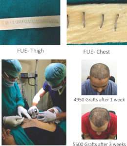
  • FUE- Painless,scarless,sutureless ?
  • Hair restoration surgery isn’t painless, but pain control is much more effective today than even in the recent past
  • Medical research has improved understanding of nociception-how a sensation of pain is received, transmitted and interpreted by nerves from the site of injury to the spinal cord and the brain.
  • There is better understanding of the relationship between effective anesthesia and limitation of bleeding or “oozing” during surgery.
  • An inadequately anesthetized, anxious patient may have increases in heart rate and blood pressure that increase risk for bleeding; thus, anesthesia level and physical signs such as heart rate and blood pressure are carefully monitored during surgery.

Medications prior to surgery

  • Preoperative anxiety about discomfort may be alleviated by administration of anti-anxiety medication.
  • Antibiotic, anti-inflammatory, pain killer should be given.

Minimising pain: Increasing comfort

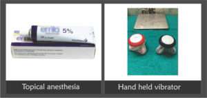
  • Topical anesthesia : EMLA cream covered with plastic 45 min-1 hr prior to surgery reduces the painful sensation of needle prick
  • Physical stimulation: We use vibrator to minimise the pain of injection
  • Melzack and Wall- Gate control theory: Touch, pressure and vibration have the potential to diminish the perception of painful stimuli.

melzack

Minimising pain: Increasing comfort

  • Needle caliber and rate, depth of injection:
  • Smaller caliber needle causes less pain than larger ones, however this potential is only realized if rate of injection is slowed, since a more rapid rate of administration increases the pain.
  • We use 30-31 G needle with slow rate of infusion.
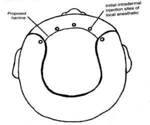
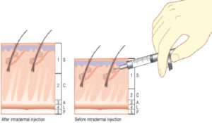
  • Depth of injection: Initial injections into deeper tissue—-less pain … and then move into superficial planes—–rapid onset and longer duration of anesthesia.
  • Office Environment: Anxious patients tend to have more intense pain
  • Prior patient education
  • Office staff demeanor
  • Light music,TV

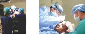
Comfortable office environment

Anaesthesia

Local anesthetic:

  • LA are divided into two categories- Ester and amide
  • In hair transplant we use amide class of anesthetic mostly Lignocaine and bupivacaine
  • As they are primarily cleared by hepatic metabolism, patients with liver disease should be treated cautiously.

Lignocaine:

  • Most common short acting LA
  • Onset of action is 1-4min and duration of action is 120 min when combined with epinephrine
  • The recommended max total daily dose (TDD) is 4.5mg/kg(max 300 mg) when used alone and 7mg/kg (max 500 mg) when combined with epinephrine.

Bupivacaine: 

  • It is the most commonly used long-acting local anesthetic in hair transplantation.
  • Onset of action is 5-10min and duration of action is 180-240 min when combined with epinephrine
  • The recommended max total daily dose (TDD) is 175 mg without and 200 mg with epinephrine

Max safe daily dose in ml

  • Lignocaine = 21.3mg/ml
  • So we can use max 500mg= 23.47ml
  • Bupivacaine= 5mg/ml
  • So we use max 200 mg= 40 ml

Lignocaine:Bupivacaine

  • Commonly bupivacaine is combined with lignocaine to achieve quick and prolonged anesthesia

Factors considered in determining max dose:

  • General health status, body weight
  • Dosing interval, length of surgery
  • Anesthetic technique
  • Administration of concomitant medications like BZD

Techniques of local anesthesia

  • Peripheral nerve block
  • Field block
  • Tumescent anesthesia

Peripheral nerve block

  • It is infiltration of small volume of LA around nerve trunks.
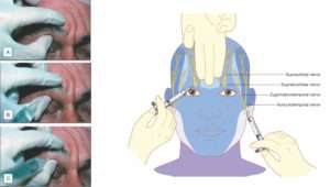
  • In hair transplant we give supraorbital,supratrochlear and occipital nerve blocks

NERVE BLOCK advantage

  • Can substitute a single injection for a large number of injections and
  • anesthetize a large area with a small amount of anesthetic so less volume of local anesthetic required

Rapid onset and prolonged anesthesia

dermatalk

(A) Technique for locating the supraorbital notch. (B) Proper position and direction of needle during infiltration of anesthetic for nerve block. (C) Needle redirected medially to block supratrochlear nerve

Field block

  • Field block involve the creation of anesthetised perimeter proximal to an operative field

Different techniques can be used to give field block are-

  • Multiple wheal technique
  • Continuous wheal technique

wheal tech

(Fig.Multiple wheal Technique)

Tumescent anesthesia

  • It employ large volume of dilute local anesthesia with adrenaline
  • It reduce local anesethetic doses, improves pain control and effectively provide hemostasis and clear operative field
  • Specially beneficial in body hair transplant where large area to be anesthetised and one can easily cross the same limit of LA

tumescent


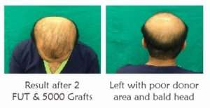
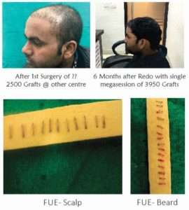
  • In previous failure cases, high grade of baldness and cases where body hair transplant is required chances of anesthesia overdose and consequent side effects are higher so thorough knowledge of anesthetic agent- its mechanism of action, dose, route, rate and depth of injection should be well versed.
  • FUT Disaster @ some other centre

22

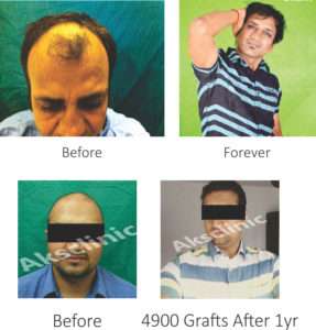
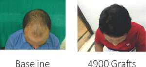
  • Correction of previous surgery- single megasession of 3950 Grafts by FUE

21

anaesthesia

Anaesthesia

cc

Conclusion

  • Anxiety about discomfort can be a roadblock. Effective patient/physician communication and rational use of medications can remove the roadblock and open the way to successful hair restoration.

Dermatological Manifestations of Chronic Kidney Disease

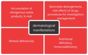
Chronic kidney disease is a progressive loss of renal function over a period of months or years , and characterized by abnormally low and deteriorating glomerular filtration rate (1). Individuals with glomerular filtration rate<60 ml/min/1.73 m2 for three monthsare classified as having chronic renal disease.End stage renal disease has highest prevalence of dermatological manifestations (50-100 % cases)

Common causes of chronic kidney disease in India are diabetes, hypertension &structural kidney defect. The prevalence of chronic renal failure is increasing in India.

Skin being an external organ is often a window to the progressive pathology going on in kidneys.

There are various skin manifestations in chronic kidney disease which can be specific or non-specific

Pathomechanism of dermatological changes-

pathomechanism

The commonly seen dermatologic manifestations are-

Cutaneous changes-xerosis, pruritus ,pigmentation abnormalities including pallor and grey-brown pigmentation,perforating disorders ,superficial infections, purpura&ecchymoses.

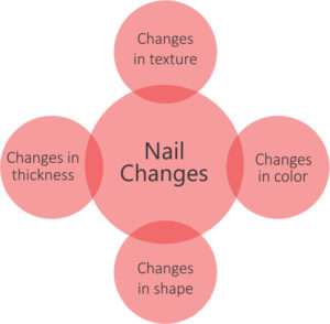
Nails –half and half nails,leukonychia,pitting,beau’s lines,splinterhaemorrhages, absence of lunula,red lunula,koilonychia

Hairs-thin and lusterless scalp hairs,telogen effluvium,hypertricosis in patients on cyclosporine.

Mucosa-xerostomia, uremic mucositis,bald tongue,pigmentation of mucosa,macroglossia with increased teeth markings,candidiasis,gingivitis,periodontitis.

The cutaneous manifestations related to hemodialysis include changes at A-V fistula site, xerosis, pruritus &pseudoporphyria

Non-specific changes-

1.Xerosis-

Xerosis is seen in 50-85% of patients of CKD.It is attributable to atrophy of sebaceous glands sudoriferous glands and volume depletion secondary to diuretics(3). Xerosiscan be treated by applying moisturizers with 5-10 % urea and 2-3% salicylic acid.

2.Pigmentary abnormalities-

a.Pallor-pallor is seen due to anemia of chronic disease

b.Yellowing of skin –yellowing of skin is due to retention of fat-soluble pigments in the dermis and subcutaneous tissue, such as carotenoids and urobilinogen

c.Grayish-brown skin-greyish brown pigmentation is seen due to the deposit of hemosiderin;

d.Hyperpigmentation associated with sun exposure

e.Ecchymosis-ecchymoses are seen secondary to platelet dysfunction

3.Pruritus

The pathophysiology of pruritus lies in xerosis,hyperparathyroidism and hypervitaminosisA,iron deficiency anemia and electrolyte imbalance.. Therapeutic modalities tried are-antihistaminics, ondansetron, serotonin antagonists,activated charcoal, cholestyramine, optimizing daily and in severe cases by narrow band UV-B & UV-A phototherapy.

4.Gynecomastia –

It is seen in males due to increase in prolactin level

5.Purpura-

They are seen secondary to defects in platelet function and use of heparin during dialysis.

6.Nail changes-

Nail changes associated with chronic kidney disease include –

a.Koilonychia-

Ridged nails that are spoon shaped & concave due to  anemia of chronic disease and decreased erythropoietin levels in chronic renal failure.

b.Absent lunula-

Absence of visible portion of white matrix. It reflects metabolic disorders  andanemia.

nail changes

Specific manifestations-

1.Perforating disorders-

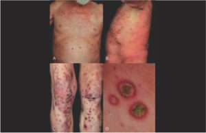
They are disorders of trans-epidermal elimination of altered keratin or dermal connective tissue material . These disorders  include- perforating folliculitis , Kyrle’s disease,elastosisperforansserpiginosa and reactive perforating collagenosis(4,5).Clinically, the lesions are hyperkeratotic to verrucouspapules ,umbilicated nodulesor plaques over extremities with central  keratotic plug. There is no effective treatment for perforating disorder,but topical retinoids,allopurinol,UVB,PUVA have been tried.

perforating

2. Calciphylaxis-

Poorly understood and highly morbid syndrome of vascular calcification and skin necrosis ;seen in stage 5 of chronic kidney disease. Lesions first present over lower extremities,abdomen,buttocksand penis as areas of mottling of skin & induration in a livedoreticularis pattern . The progression is very rapid leading to formation of ulcerated,gangrenous lesions.Prognosis is very poor in untreated  patients with  ulceration , with mortality rate of 60-80 %. It can be treated by cincalcet,sodium thiosulphate and surgical debridement for ulcerated lesions.

3. Nephrogenic systemic fibrosis (NSF)-

Scleroderma like changes in skin and internal organs in patients of CRF exposed to gadolinium based contrast agents.

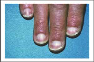
4. Half and half nails-

It is characterized by a proximal whitish band and distal brownish or pinkish discoloration. The pathogenesis is attributed to an increase of the melanocytic stimulating hormone and increase in capillary number and thickness.

half and half nails

5. Uremic frost-

One of the rarest presentations is uremic frost, seen in only 3% of the patients with ESRD. It is related to frank uremia and recognized as a white deposit on the skin, secondary to crystallized urea excreted in sweat.

6. Bullous disease of dialysis –

Bullous lesions on photoexposed parts of end-stage renal failure resembling lesions of porphyria cutaneatarda.The porphyrin levels are normal or mildly raised.

7. Arterial steal syndrome –

The arterial steal syndrome is an uncommon but highly morbid complication of the vascular access necessary for hemodialysis. Presents as Pale/blue and/or cold hand without pain/or pain during exercise and/or hemodialysis, ischemic pain at rest, ulceration, necrosis and gangrene.

8. Uremic stomatitis-

It clinically represents as white plaques distributed predominantly on the buccal mucosa, floor of the mouth and tongue(6).It is considered as the reaction to a tissue irritant, possibly ammonia compounds derived from urea hydrolysis by salivary urease, whenever salivary urea level exceeds 180gm/dl. Treatment with a mildly acidic mouth rinse,  such as diluted hydrogen peroxide clears oral lesionsUremic stomatitis

Conclusion:

Skin manifestations are fairly common and troublesome to patients affected by chronic renal failure. They negatively affect the quality of life of these patients causing constant worry in health care team. Therefore it is essential to identify and treat these diseases at the earliest.

References-

  1. Johnson AC, Leway AS, Coresh J, Levin A, Lau J, Eknoyan G: Clinical practical guidelines for Chronic Kidney Disease in adults: Part I. Definition, Disease stages, Evaluation, Treatment, and Risk factors. Ameri­can Family Physician 2004;70:869-876.
  2. Prabahar MR, Chandrasekaran V, Soundararajan P. Ep­idemic of chronic kidney disease in India—what can be done? Saudi J Kidney Dis Transpl 2008;19:847-53.
  3. Udayakumar P, BalasubramanianS,Ramalingam KS et al. Cutaneous manifestationsin patients with chronic renal failure onhemodialysis. Indian J DermatolVenereolLeprol2006; 72: 119-25.
  4. Maurice PDL. Acquired perforating dermatosisin renal patients.Nephrol Dial Transplant.1997;12:2774-5.
  5. Lynde CB, Pratt MD. Acquired perforating dermatosis: association withLynde CB, Pratt MD. Acquired perforating dermatosis: association with diabetesand renal failure.CMAJ. 2009;181:615
  6. Neville BW, Damm DD, Allen CM. Oral manifestation of systemic diseases. In: Bouquot JE (Ed). Oral and Maxillofacial Pathology (2nd ed). USA: WB Saunders Company 2002:705-36