DERMASCOPE -IS IT WORTH THE INVESTMENT?

Introduction

The term “dermoscopy,” first coined by Friedman is also known as dermatoscopy, epiluminescence microscopy, or surface microscopy. It is a simple, noninvasive and practical technique in which a dermoscope, a handheld device with 10x magnification, is used to examine skin. Aptly called as “The dermatologist’s stethoscope”, it is a relatively young, dynamic and exciting field. 1It allows rapid in vivo evaluation of morphologic structures of epidermis, dermoepidermal junction and papillary dermis which cannot be seen by naked eye.3,4 Since the dermoscopic structures correlate well with the histopathologic alterations, dermoscopy represents an interface between clinical and histopathological findings.1,3

Historical aspects

Dermoscopy was first introduced for evaluation of pigmented / non-pigmented skin tumors. Earlier dermatoscopes used in late 1980’s were large devices that required an oil or alcohol interface to reduce light reflection, later in 1990s hand-held devices started being used. 2

Principle of dermatoscope

When a skin lesion is visualized with a magnifying loupe with lighting, most of the light is scattered due to reflective property of stratum corneum. In order to allow more light to pass through stratum corneum, to enable deeper visualization of pigment and vascular patterns, two ways can be used. One is by using a fluid medium as an interface and a transparent glass contact plate and the other is by using a cross polarized light.The newer dermoscopes are provided polarized light allowing both contact and noncontact dermoscopy.2

 Dermatoscopes available

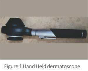
  1. Handheld polarized dermoscopes

They are highly portable with magnification of 10 to 20 X, do not require skin contact or immersion fluid and allow rapid and safe examination without the risk of possible transfection .3,4These devices can distort colors, so they also have contact dermoscopy capabilities to minimize color distortions.1 They can be used to take photographs by connecting them with cameras, smart phone cameras and digital SLRs, using adapters.Figure 1 shows a hand held dermatoscope.

dermatoscope

  1. Nonpolarised light contact dermoscopy

In contact dermoscopy, the device emits a beam of light which strikes the skin at a 20º angle. To eliminate reflections and thus enable better visualization, a fluid like oil, water, gel, glycerin, or ethanol gel is placed between the skin and the device.1 These devices  are not preferred as they may reduce the vessels (due to pressure) and/or scaling (when using a liquid interface) visibility, but some superficial findings like comedo-like structures, milia-like cysts and crypts (seen in seborrheic dermatitis) and the blue-white veil seen in melanoma are better seen with these devices.5 Disposable contact caps, food wraps, adhesive tapes, and 70% ethanol (as interface fluid) help to avoid cross infection in contact dermoscopy.2

  1. Video dermoscopes

These devices need to be connected to a viewing monitor, usually with a USB connection. Advantages are that a larger view is obtained which can be shared with the patients/ dermatologists and images can be stored. These devices are expensive and are usually restricted to research centers.2 

  1. Multispectral dermoscopes and UV dermoscopes

These allow better visualization of specific structures eg surface pigmentation is better seen under blue light, superficial vascularity under yellow light and deeper pigmentation and vascularity with red light which penetrates deeper.2

A good quality hand-held, portable device with both contact dermoscopy and cross polarized light options can be considered best for a beginner.2

Uses of dermatoscope4,5

Dermoscopy is useful for diagnosis of common dermatologic diseases, especially in atypical/unusual cases. 3Various uses of dermoscopy include

1. Dermoscopy for pigmented and non-pigmented skin tumors including melano­cytic/ non-melanocytic and benign/ malignant skin tumors.

a.Dermoscopy provides useful and reliable information on the histopathologic basal cell carcinoma (BCC) subtype, reveals tumor characteristics like ulceration or pigmentation, facilitating the accurate management of the tumor. Presence of ulcerations represents a predictor of favorable response to imiquimod. Dermoscopy reveals pigmentation in 30% of clinically non-pigmented BCCs, which is relevant for BCCs scheduled to be treated with photodynamic therapy (PDT), as pigmented tumors are less responsive to PDT.3

b.Dermoscopy allows accurate assessment of true extension of  tumor,  facilitating a more  precise estimation of the required surgical margins, thus  helping to minimize  recurrence rates.3

c.Dermoscopy is useful for diagnosing different stages of keratinocyte skin cancer, thus improving the treatment.3

2. Inflammoscopy for various inflammatory diseases like psoriasis, lichen planus, pityriasis rosea etc.

Because of specific features on dermoscopy, the differential diagnosis of the papulosquamous disorders becomes simpler, permitting recognitation of lichen planus (Wickham striae), eczema (yellow crusts and patchy dotted vessels), and pityriasis rosea (peripheral white scales). The inflammatory dermatoses may be a major reason for use of dermatoscope given their incidence and the difficulties in their differential diagnosis.3

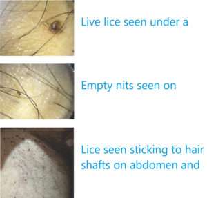
3. Entomodermoscopy for skin infections and infestations caused by parasites, viral, bacterial, fungal or protozoan infections.

Both common (warts, molluscum contagiosum, mycosis and mite infestations) and uncommon (myiasis and tinea nigra) infestations can be diagnosed by dermoscopy, scabies representing a good example showing the delta-wing jet with contrail.3

4. Trichoscopy for diagnosing hair and scalp disorders.

The term was coined by Rudnicka and Olszewska in 2006. Trichoscopy improves diagnostic accuracy and may help in better understanding of pathogenesis of hair disorders.6

5. Nail/nailfold abnormalities (Onychoscopy/ Capillaroscopy) 7

a.It may be useful for diagnosis of small, subclinical/ sub­ungual warts or for the assessment of therapeutic response after treatment.Decrease of capillary density within viral warts is observed in lesions responding to treatment.

b.Dermatoscopy helps to localize tumors and to visualize vascular patterns. It facilitates the delimitation of surgical margins before treatment.

c.In subungual melanoma, the Hutchinson sign defined by the visibility of a pigmentation of the periun­gual cuticula, can be demonstrated by dermatoscopy.

d.Sequential digital dermatoscopy may help to detect dynamic changes in color and increase in width of whole longitudinal pigmentation over a period of time.

e.Nail fold capillaroscopy is useful for screen­ing of autoimmune diseases. Presence of dilated capillaries and/or nail fold hemorrhage on dermatoscopy is a sensitive and specific for diagnosis of systemic sclerosis.

6. Other disorders

a.The detection of a yellow-orange background is considered a dermoscopic clue for the diagnosis of granulomatous skin disorders like sarcoidosis, lupus vulgaris.3

b.Dermoscopy has been found useful in the diagnosis of rosacea, discoid erythematous lupus, morphea, lichen sclerosus, pigmented purpuric diseases, Darier’s disease, Grover’s disease, porokeratosis  etc.3

7. Dermoscopy for monitoring.

a.Utility of dermoscopy for therapeutic monitoring of imiquimod treatment has  been reported.1

b.Dermatoscopy has been used to evaluate vitiligo activity. Balakrishna et al used dermatoscope to monitor the result of various surgical procedures in vitiligo.8

Criteria/factors to be considered while using dermoscopy2,5  

The criteria include

  1. Morphology/arrangement of vascular structures
  2. Scaling patterns
  3. Colours
  4. Follicular abnormalities
  5. Specific features

The factors include

  1. Quality
  2. Budget
  3. Need for documentation
  4. Major types of cases one sees

Advantages of dermatoscopy 1

  1. It reduces the uncertainty of clinical examination, thus improving diagnostic accuracy.
  2. It enables detection of tumors at earlier stages, thus improving the prognosis.
  3. It reduces the need for unnecessary biopsies.
  4. It provides guidance on the optimal site for biopsy, thus improving the histopathological accuracy.

Studies on dermatoscopy

Scientific publications on dermoscopy have  increased significantly with more than  1000 articles  been published between 2003 and 2007.9 Criteria and dermoscopic patterns have been established for melanoma /melanocytic nevi, basal cell carcinoma, seborrheic keratosis, squamous cell carcinoma, Bowen’s disease and many more benign skin tumors. Trichoscopic criteria for localized and diffuse hair loss have also been described.2

International Dermoscopy Society

The International Dermoscopy Society (IDS) founded in 2003, promotes education and research, and conducts conferences and multicentre studies in the field of dermoscopy.2,9

Conclusion

Dermoscopy is an irreplaceable, noninvasive and helpful auxiliary tool in assisting the recognition/ differential diagnosis of various dermatoses, reflecting their histopathological background, thus having a great impact on clinical practice of dermatology.5Thus it is definitely worth the investment.

References

  1. Heliane Sanae Suzuki,Sérgio Zuñeda Serafini, Maurício Shigueru Sato. Utility of dermoscopy for demarcation of surgical margins in Mohs micrographic surgery.An Bras Dermatol. 2014 Jan-Feb; 89(1): 38- 43.
  2. Feroze Kaliyadan.The scope of the dermoscope. Indian Dermatol Online J. 2016 Sep-Oct; 7(5): 359–363.
  3. Teresa Russo, Vincenzo Piccolo,Aimilios Lallas, Giuseppe Argenziano. Recent advances in dermoscopy. Version 1. F1000Res. 2016; 5: F1000 Faculty Rev-184.
  4. Iris Zalaudek, Aimilios Lallas, Elvira Moscarella,, Caterina Longo,  H. Peter Soyer, Giuseppe Argenziano. The dermatologist’s stethoscope- traditional and new applications of dermoscopy . Dermatol Pract Concept 2013;3(2):11
  5. Enzo Errichetti . Giuseppe Stinco. Dermoscopy in General Dermatology: A Practical Overview. Dermatol Ther (Heidelb) (2016) 6:471–507.
  6. Vivek V Nikam , Hita H Mehta.A Nonrandomized Study of Trichoscopy Patterns Using Nonpolarized (Contact) and Polarized (Noncontact) Dermatoscopy in Hair and Shaft Disorders. Int J Trichology. 2014 Apr-Jun; 6(2): 54–62.
  7. Holger A. Haenssle, Andreas Blum, Rainer Hofmann-Wellenhof, Juergen Kreusch, Wilhelm Stolz, Giuseppe Argenziano, Iris Zalaudek, Franziska Brehmer. When all you have is a dermatoscope-start looking at the nails.  Dermatol Pract Concept 2014;4(4):2
  8. Balakrishnan Nirmal.Use of Dermatoscope to Monitor the Repigmentation of Various Vitiligo Surgical Procedures. J Cutan Aesthet Surg. 2016 Oct-Dec; 9(4): 286-287.
  9. Giuseppe Argenziano, Iris Zalaudek. Dermoscopy: a new perspective. Dermatol Pract Concept. 2011 Jan; 1(1): 57-58.

Polycystic Ovarian Syndrome: Clinical Guidelines    

The lack of awareness of PCOS among patients and health care professionals made 70% of women with PCOS remain undiagnosed- Australian study1

Introduction

Polycystic ovary syndrome (PCOS) is the most common endocrinological abnormality in patients with acne with an incidence of 5–10%, Around 30% of all patients with PCOS show clinical features of acne together with clinical or laboratory aspects of hyperandrogenism, oligo-amenorrhoea and ovarian cysts.2 Studies of PCOS in India carried out in convenience samples reported a prevalence of 3.7% to 22.5%, with 9.13% to 36% prevalence in adolescents only.

There is a high incidence of psychological issues amongst women with PCOS such as depression, anxiety and eating disorders when compared to the general population. A woman with PCOS may need to see a general practitioner, dietician, dermatologist, psychologist, exercise physiologist, endocrinologist, gynaecologist and infertility specialist.

Pathophysiology of acne and hyperandrogenism in PCOS

Before puberty, the adrenal glands produce increasing amounts of dehydroepiandrosterone sulphate (DHEAS), which can be metabolized into more potent androgens in the skin, driving enlargement of the sebaceous gland and increased sebum production; this is a prerequisite for acne in all populations.

Molecular aspects

Sebocytes and keratinocytes of the pilosebaceous follicle infundibulum in patients with acne have androgen receptors that are both more numerous and more sensitive than those in normal subjects.3,4 The androgenic activity may be induced by potent androgens, such as testosterone and dehydrotestosterone (DHT) and weak androgens, such as dehydroepiandrosterone (DHEA) and androstenedione, which reach the skin through the bloodstream or can be produced locally on the skin. In sebocytes and keratinocytes of the infundibulum the enzyme 5-alpha reductase type 1 can transform testosterone into DHT and this enzyme activity is greater in the follicular infra-infundibulum than in the epidermis. DHT acts 5–10 times more strongly than its precursor testosterone.

Clinical aspects

PCOS patients can present with any one or in combinations with various clinical features such as hirsutism, female-pattern androgenetic alopecia, irregular menstrual periods, obesity, deepening of the voice and clitoridomegaly. Infertility, diabetes, insulin resistance and cardiovascular disease can also be associated with PCOS.

Androgen excess is often characterized by nodulocystic acne, dispersed rapidly growing comedones and severe, sudden-onset acne. It is wise to remember that many women with androgen excess can still have a normal menstrual period.5, 6

Diagnosis

The Rotterdam 2004 Consensus Workshop (Revised 2003) recommended that two of the following criteria should be present to establish the diagnosis of PCOS:

1) Chronic oligo or anovulation for more than 6 months

2) Clinical and/or biochemical evidence of hyperandrogenism, and

3) Polycystic ovaries in ultrasound.

Other disorders that mimic PCOS phenotype should be excluded such as thyroid dysfunction, congenital adrenal hyperplasia, hyperprolactinaemia, androgen-secreting tumours and Cushing’s syndrome.


Androgen assessment in women generally remains controversial. Calculated bioavailable testosterone and calculated free testosterone can be used interchangeably as a marker of testosterone. The FAI (100 x (total testosterone/SHBG)) is also a widely used measure of androgens. Calculated bioavailable testosterone, calculated free testosterone or free androgen index should be first line investigation; androstenedione and dehydroepiandrosterone sulfate could be used as second line investigation for biochemical determination of hyperandrogenism in polycystic ovary syndrome.


Levels of androgens outside the expected range can also be indicative of other diseases like, high levels of testosterone (> 150– 200 ng/dL), in particular if associated with normal levels of DHEA, are suggestive for ovarian tumour, high levels of DHEA (> 8000 ng/mL) are suggestive for adrenal tumours. Slightly elevated levels of DHEA (4000–8000 ng/mL) are found in CAH, PCOS and Cushing disease. A ratio of LH/FSH greater than 2: 1 is indicative of a possible androgenism such as in PCOS.

Blood levels of Prolectin largely above the normal values may be related to a prolactinoma and a head scan, preferably a MRI, should be performed. High levels of 17-OHP and a positive adrenocorticotropic hormone (ACTH) stimulation test are essential to make the diagnosis of CAH. The best time to run laboratory tests is within the first week of the menstrual period.

If biochemical hyperandrogenism is needed for the diagnosis of polycystic ovary syndrome, the oral contraceptive pill could be withdrawn for three months to facilitate appropriate hormonal assessments. However, the risk of unplanned pregnancy needs to considered and weighed up against potential benefits of early diagnosis. Contraception may still need to be otherwise managed during this time.


Given the apparent lack of specificity of polycystic ovaries on ultrasound in adolescents, generally, ultrasound should not be recommended first line and if pelvic ultrasounds are to be ordered in adolescents, the results should be interpreted with caution.


Testing of fasting insulin and lipids should be requested in patients with PCOS due to the metabolic consequences that occur more frequently in these cases.

Biochemical-hyperandrogenism

Insulin levels should be measured in overweight and obese patients and those with a family history of diabetes and not responding to treatment.

Those women with polycystic ovary syndrome should be considered at increased relative risk of Dyslipidemia and cardiovascular disease (obesity, cigarette smoking, dyslipidemia, hypertension, impaired glucose tolerance, lack of physical activity). To assess for risk of type 2 diabetes, in PCOS patients, age, gender, parental history of diabetes, hypertension, waist circumference, high GTT should be assessed.

Treatment

Hormonal treatment is indicated as first line of treatment in cases of papulopustular, nodular and conglobate acne in females with identified hyperandrogenism, in adult women who have monthly flare-ups and when standard therapeutic options are unsuccessful or inappropriate.PCOS. In comedonal and mild-to moderate papulopustular acne hormonal treatment is not recommended. It can take 3–6 months to see clinical improvement, although effects may be seen after one menstrual cycle.

Hormonal therapy

Hormonal therapies that can be considered for patients with acne include: COCs; anti-androgens like Cyproterone acetate (CPA), spironolactone, flutamide; and GnRH agonists. Metformin is not strictly classified as a hormonal therapy for acne, but it may be of help in some cases of hyperandrogenism with hyperinsulinaemia whether or not the patient is overweight.

COCs- The most commonly used oestrogen is ethinylestradiol (EE) and group of progestins includes CPA, chlormadinone and drospirenone, and derivatives of 19-nortestosterone (estranges and gonanes) like norethindrone, levonorgestrel, norgestrel, desogestrel, norgestimate and gestodene.7-10

Guidelines:

The derivatives of 19-nortestosterone interact with progesterone receptors and also cross react with androgen receptors, resulting in an androgen-like effect with possible worsening of acne.

COCs containing chlormadinone acetate or CPA performed better than levonorgestrel, CPA showed better acne outcomes than desogestrel; levonorgestrel performed slightly better than desogestrel; and a drospirenone-containing COC showed more effectiveness than norgestimate or nomegestrol acetate but less effectiveness than CPA in acne outcomes.

Several safety issues are related specifically to the use of oestrogens in COCs: breast tenderness, headache, nausea, bloating, hypercoagulability, increased cardiovascular disease, increased risk of endometrial and breast cancers. The use of COCs comes with several safety considerations. Venous thromboembolism (VTE) and arterial thromboembolism are the most serious potential side-effects associated with COCs. Recent formulations of COCs with low dosages of EE (20–35 lg and 15 lg in some cases) reduce the risk of oestrogen-related side-effects.

CPA is a progestin that is used in combination with an oestrogen in the majority of the cases (such as in COCs) but sometimes it is prescribed alone. When used alone, at the recommended dose of 50–100 mg daily, CPA treatment may be initiated from the first or fifth day of the menstrual period and stopped on the 14th day just before ovulation. Side-effects are menstrual irregularities (amenorrhea, oligomenorrhea), breakthrough bleeding, breast tenderness, headache and nausea, although they tend to regress with time.

Spironolactone is a synthetic steroidal derivative of aldosterone that works as an aldosterone antagonist. Standard doses are 50–100 mg once or twice daily, taken with meals, with a steady-state daily dose range between 25 and 50 mg. Side-effects including menstrual irregularities, breast tenderness or enlargement are more common at higher doses, but usually they are mild in severity and a reduction of the dosage is sufficient to reduce them to acceptable levels. Spironolactone is contraindicated in pregnancy (FDA pregnancy category C) because of a potential for feminization of the male foetus and the use in combination with a contraceptive pill is advisable and recommended. The dose is titrated from 25 mg, with an increase of 25 mg every 20 days until the maximum dose of 100 mg has been reached, according to tolerability and blood levels of potassium.

Anti-androgen activity of Flutamide is mostly related to the inhibition of androgen receptors, in particular the ones that bind DHT. Flutamide can cross the placental barrier and cause a pseudohermaphrodite condition in a male fetus; it cannot be given to pregnant women. The standard dose used in acne ranges between 625 and 500 mg daily, which can result in an 80% improvement of acne.

Metformin causes decrease in insulin (about 20-30%) and testosterone (about 20%) with increase in SHBG. Metformin should not be kept as first line of treatment in adolescent and unmarried females with regular menses and devoid of anovulatory cycles. Metformin could be used alone to improve ovulation rate and pregnancy rate in women with polycystic ovary syndrome who are anovulatory, have a body mass index ≤30kg/m2 and are infertile with no other infertility factors. For treatment of infertility a referral to gynaecology should be made for ovulation induction.


Weight loss should be targeted in all women with polycystic ovary syndrome and body mass index ≥25kg/m2 (overweight) through reducing dietary energy (caloric) intake in the setting of healthy food choices, irrespective of diet composition. Exercise participation of at least 150 minutes per week should be recommended to all women with polycystic ovary syndrome, especially those with a body mass index ≥25kg/m2 (overweight), given the metabolic risks of polycystic ovary syndrome and the long term metabolic benefits of exercise. Of this, 90 minutes per week should be aerobic activity at moderate to high intensity (60% – 90% of maximum heart rate) to optimise clinical outcomes.

PCOS

Mechanism of action of hormonal treatment for PCOS


References

  1. March, W., et al., The prevalence of polycystic ovary syndrome in a community sample assessed under contrasting diagnostic criteria. Human Reproduction, 2010. 25(2): p. 544-51.
  2. Bettoli et al., Is hormonal treatment still an option in acne today. BJD, 2015: p. 37-46.
  3. Lai JJ,Chang P, Lai KP et al. The role of androgen and androgen receptor in the skin-related disorders. Arch Dermatol Res 2012; 304:499–
  4. Chen W,Thiboutot D, Zouboulis CC. Cutaneous androgen metabolism: basic research and clinical prospectives. J Invest Dermatol 2002; 119:992–
  5. Darley CR,Moore JW, Besser GM et al. Androgen status in women with late onset or persistent acne vulgaris. Clin Exp Dermatol 1984; 9:28–
  6. Thiboutot D.Acne: hormonal concepts and therapy. Clin Dermatol 2004; 22:419–
  7. Thiboutot D,Archer DF, Lemay A et al. A randomized, controlled trial of a low-dose contraceptive containing 20 microg of ethinyl estradiol and 100 microg of levonorgestrel for acne treatment. Fertil Steril 2001; 76:461–8..
  8. Leyden J,Shalita A, Hordinsky M et al. Efficacy of a low-dose oral contraceptive containing 20 μg of ethinylestradiol and 100 μg of levonorgestrel for the treatment of moderate acne: a randomized, placebo-controlled trial. J Am Acad Dermatol 2002; 47:399–409.
  9. Maloney JM,Arbit DI, Flack M et al. Use of a low-dose oral contraceptive containing norethindrone acetate and ethinyl estradiol in the treatment of moderate acne vulgaris. Clin J Women’s Health 2001; 1:123–
  10. Lucky AW,Henderson TA, Olson WH et al. Effectiveness of norgestimate and ethinylestradiol in treating moderate acne vulgaris. J Am Acad Dermatol 1997; 37:746–54.

Pediculosis Corporis: Really a Disease of the Dirty?

Pediculosis corporis, also known as Pediculosis vestimenti and Vagabond’s disease is a cutaneous condition caused by body lice (Pediculus corporis) that lay their eggs in the seams of clothing.


Pediculosis pubis is usually sexually transmitted, and can extend beyond the pubic area to involve other areas of the body, including the eyelashes (pediculosis ciliaris). Phthirus pubis, also known as the crab louse, is the responsible organism.


Body lice frequently lay their eggs on or near the seams of clothing. They must feed on blood and usually only move to the skin to feed. They exist worldwide and infest people of all races. They spread rapidly under crowded living conditions where hygiene is poor through prolonged direct physical contact or through contact with fomites like linens or towels. Infestation is unlikely in anyone who bathes regularly and who has at least weekly access to freshly laundered clothing and bedding.


A 58-year old diabetic man presented with severe itching over abdomen, pubic area and upper thighs for 6 weeks. On examination, black dots were seen adhering to the hair shafts on abdomen and upper thighs. On closer examination, these dots were found to be lice. A dermatoscope helped in magnifying lice stuck to the hair shaft and also in documenting the movement. He was diagnosed as a case of pediculosis corporis. Patient denied history of same in any other family member although he was requested to bring his wife for examination. The patient and his clothes were neat in appearance, in contrast with the usual presentation of the disease. He was given oral ivermectin in dose 200mcg/kg stat dose and to be repeated after 2 weeks. He was also prescribed weekly application of permethrin 1% cream for half an hour for 2 weeks and anti histamines to be taken as required. The importance of personal hygiene was repeatedly emphasised. He was advised washing all clothes and bedding in boiling water thoroughly. He was asked to leave other fomites which could not be cleaned in this manner untouched and isolated for a week by which the louse would have died in absence of nutrition. His follow up visit after 2 weeks showed almost complete clearance of body lice, but empty nits were still seen adherent to the hair shafts. He was advised to shave the affected area. His blood sugar levels were under control throughout. He was advised HIV testing but the report is yet to be seen.


This case is interesting as pediculosis corporis is traditionally seen as a skin condition affecting persons with poor hygiene while this patient who had impeccable hygiene has a florid presentation in absence of any immune suppression.

pediculosis

DISSEMINATED Histoplasmosis in an Immunocompetent Female

Case Report:

A 40year old housewife presented with the complaints of recurrent episodes of high grade fever from 3 months and asymptomatic skin colored papular eruptions over the central face for last 1 and 1/2 month. Subsequently, the similar papules had appeared over chest, abdomen, back and upper limbs. There was no history of breathlessness, cough, seizures and altered sensorium. Patient was a known diabetic and was on oral hypoglycemic drugs. There was no history of extramarital sexual exposure and blood transfusion. No history of travel beyond nearby locales.  Clinical examination revealed that the patient was pale and had hepatospleenomegaly. Multiple skin colored to erythematous discrete as well as coalescing papulonodular lesions ranging in size from 0.2 cm to 1cm present bilateral symmetrically over the central part of the face involving the glabella, medial side of both the eyebrows, nasolabial folds, chin, neck, anterior chest,  abdomen, back  and both cubital fossa. Few of these papules showed central umblication and central necrosis with haemorrhagic crusts. Oral mucosa was also involved with multiple superficial erosions over hard palate and both buccal mucosa. Patient had generalized lymphadenopathy , lymph nodes were firm, non-tender, mobile, painless with no matting or overlying sinuses. A review of the central nervous system, respiratory system and cardiovascular system revealed no evident abnormality.

Further, the patient was admitted and subjected to a battery of  investigations:

  1. Haematological Studies: Haemoglobin was 8.4 mg%, Total leucocyte count was 8000/μl and platelet count was 2.8 lakhs/μl at the time of admission but these parameters deteriorated with time to Hb- 6 gm%, and TLC- 6000/μl while platelet count was unaltered. The DLC- P73L26E1. On peripheral blood smear the RBCs were normocytic normochromic with few macrocytic normochromic cells, WBCs shows normal morphology while platelet count were adequate. Her corrected reticulocyte count was 1.5%. Her absolute lymphocyte count was 700/μl and CD4 T helper cell count was 289/μl and CD4/CD8 ratio was 1.8.
  2. Biochemical Investigations: Her random blood sugar was 244 mg%. On LFT, Serum bilirubin (Total)- 2.9mg% while bilirubin(Direct)- 0.9mg%, SGOT levels were 231U/l, SGPT- 6 U/l and Alkaline phosphatase levels were 1460U/l. The total protein levels were 6.2 gm%, serum albumin – 2.5gm% and serum globulin of 3.7 gm%. Her Na+ levels were 138 meq/l and K+ levels were 4.2 meq/l at the time of admission but on treatment with Amphotericin her K+ levels were reduced to 2.8 meq/l. Her HIV status was negative for both HIV 1 and 2. The KFT, Urine routine microscopy, coagulation profile was within normal limits.
  3. Radiological Investigation: On USG abdomen her liver was 17.8cm with grade II fatty changes while her spleen was 4.3cm with no free fluid in chest and abdominal cavity at the time of admission. The plain chest Xray PA view showed bilateral nodular opacities. The NCCT head showed bilateral periventricular white matter and centrum semiovalehypodensities suggestive of ?Ischaemia ??Infection while rest of the brain was normal.
  4. Histopathology: The skin biopsy from the papular lesion showed thinned and flattened epidermis. The dermis shows sheets of foamy macrophages containing numerous basophilic dot like inclusion surrounded by a clear halo. The PAS positive bodies were present. All these findings were highly suggestive of cutaneous histoplasmosis.
  5. Tissue Culture: On Sabouraud’s dextrose agar showed colonies ofHistoplasma capsulatum
  6. Bone marrow biopsy: The spaces were occupied by cellular bone marrow which comprised of myeloid and erythroid series cells. Adequate megakaryocytes seen showing normal morphology. Histiocytes are seen showing intracellular round to oval structures and these were PAS positive. The above findings suggestive of histoplasma infection involving bone marrow.

Treatment:

Thus, a disseminated histoplasma infection was confirmed and patient was admitted started on I/V Amphotericin infusions(0.5- 1 mg/kg/d) given over 2hrs daily for 7 days. However, patient developed pedal edema and swelling of her body alongwith high grade fever, breathlessness and altered sensorium after 1 week of therapy and and unfortunately succumbed to her disease within 10 days after presentation.  Meanwhile, the underlying immunosupressed status except for diabetic status could not be ascertained.

Discussion:

Histoplasmosis, a systemic mycosis caused by the dimorphic fungus Histoplasma capsulatum var capsulatum and Histoplasma capsulatum var duboisii is endemic to many parts of the world. The clinical manifestations range from acute or chronic pulmonary infection to a progressive disseminated disease. After initial exposure to the fungus, the infection is self-limited and restricted to the lungs in 99% of healthy individuals. The remaining 1%, however, progress to either disseminated or chronic disease involving the lungs, liver, spleen, lymph nodes, bone marrow or rarely, the skin and mucous membranes. Most patients who develop disseminated histoplasmosis are immunosuppressed (eg, AIDS, solid organ transplantation, treatment with tumor necrosis factor-alpha inhibitors) or are at the extremes of age. Male gender, smoking, alcoholism, prolonged permanence in a rural environment, with exposure to bats in caves (revealing the occupational risk involved in agricultural and animal husbandry activities), houses, and holes dug in the ground are risk factors commonly found in patients with the chronic pulmonary and subacute/chronic disseminated forms of histoplasmosis.  Acute disease is usually a localised pulmonary infection, more common in children after acute exposure to the excrements of birds and cattle. Such patients achieve clinical cure spontaneously, but 58.9% require antifungals. In patients with localised disease or with some dissemianation, Itraconazole is considered as highly effective while in disseminated form Amphoterecin b is recommended.

Despite various investigation, no evidence of underlying immunosuppression was found in our patient.

References:

  1. Faiolla Rivian Christina Lopes, Coelho Mariana Correa, Santana Rodrigo de Carvalho, Martinez Roberto. Histoplasmosis in immunocompetent individuals living in an endemic area in the Brazilian Southeast. Rev. Soc. Bras. Med. Trop.  [serial on the Internet]. 2013  Aug [cited  2014  Nov  21] ;  46( 4 ): 461-465. Available from: http://www.scielo.br/scielo.php?script=sci_arttext&pid=S0037-86822013000400461&lng=en.  http://dx.doi.org/10.1590/0037-8682-0124-2013.
  2. Etxeberria-Lekuona D, Hurtado Ilzarbe G, Méndez-López I, Pérez Ricarte S, Acha Arrieta V. Acute progressive disseminated histoplasmosis in a young immunocompetent patient. Rev Clin Esp 2013;213(9):95-6.
  3. Sharma S, Kumari N, Ghosh P, Aggarwal A. Disseminated histoplasmosis in an immunocompetent individual-a case report.Indian J Pathol Microbiol2005;48:204–6.
  4. Alcure ML, Di Hipólito Júnior O, Almeida OP, Bonilha H, Lopes MA. Oral histoplasmosis in an HIV-negative patient.Oral Surg Oral Med Oral Pathol Oral Radiol Endod2006;101:33–6.

Histoplasmosis

Ochronosis a Rising Concern for Dermatologists

Exogenous Ochronosis (EO) is avoidable dermatitis, on the rise cause of unjustified use of topical hydroquinone; it is mainly encountered in melasma patients on triple therapy and is turning out to be a menace cause of difficulty in its treatment. Phenol is also known to a cause the condition.

Pathologically, a microscopic deposition of yellow (ocher-colored) pigment is found in the dermis. It is clinically and histopathologically similar to endogenous Ochronosis, also known as alkaptonuria; difference between them is that it does not present systemic manifestations such as history of dark urine, gray-blue or gray-brown coloration of cartilages, conjunctive and arthropathy and the affected area is limited to that treated with HQ.

In 1912Berddard and Plumtre described an EO case in a patient that had used phenol to treat a lower limb ulcer.In 1975 Findlay described the first EO case related to HQ,

Frequently used for the treatment of melasma, hydroquinone interferes in skin pigmentation through alteration of melanin formation, interaction with copper at the site of tyrosinase activity and in inhibition of DNA and RNA synthesis.

Hydroquinone in hydro alcoholic solution, which has a significantly higher penetration in the skin, is also an important contributing factor for EO development.

GRADING

In 1989, Hardwick et al.[41] in their epidemiological study based on clinical diagnosis graded EO as:

  • Grade I – Faint macular sooty pigmentation
  • Grade II – Distinct macular stippling/small papules
  • Grade III – Dark deposits and papules
  • Grade IV – Colloid milia (1 mm and greater) [Figure 4]
  • Grade V – Keloid‑like nodules and cysts.

SYMPTOMS

  • yellow-brown, banana-shaped fibers
  • caviar-like papules
  • brown-grey or blue-black hyper-pigmentation

The majority of the lesions will be seen on areas of the body that get the most sun.

SYMPTOMS

Pathogenesis of Ochronosis

Ochronosis

TREATMENT

Exogenous Ochronosis is not easy to treat therefore prevention is extremely important.

  • Avoiding the causative agent of the disease.
  • The use of lower HQ concentrations.
  • Sun protection.
  • Early diagnosis of irritation.
  • Suspension of treatment in case there is no clinical improvement within six months.

Treatment modalities

  • Sunscreens are extremely beneficial in limiting further augmentation of the condition.
  • Glycolic acid peelings.
  • Retinoic acid gives appreciable improvement.
  • Low potency corticosteroids in association with photo protection.
  • Dermoabrasion.
  • CO2 laser and Q-switched laser.

However, the results are not uniform and this condition continues to be of difficult treatment.

Hence, Hydroquinone prescription should be associated with full knowledge of side effects and orientation about proper use, to prevent the occurrence of this cosmetically disfiguring condition.

REFRENCES

  1. Tan SK, Sim CS, Goh CL. Hydroquinone-induced exogenous ochronosis in Chinese – two case reports and a review. Int J Dermatol. 2008;47:639-40.
  2. Online Etymology Dictionary. Available from :http://www.etymonline.com. [Last accessed on 2013 oct 06].
  3. Longo N. Inherited disorders of amino acid metabolismpresenting in adults. In: Braunwald E, Fauci AS, Kasper DL, Hauser SL, Longo DL, Jameson JL, editors. Harrison’s Principles of Internal Medicine. 15th ed. New York: McGraw Hill; 2001.p. 2307.
  4. Zawar VP, Mhaskar ST. Exogenous ochronosis following hydroquinone for melasma. J CosmetDermatol. 2004;3:234-6.
  5. Fisher AA, Davis MW. Alkaptonuricochronosis with aortic valve and joint replacements and femoral fracture: A case report and literature review. Clin Med Res 2004;2:209‑ Laskar FH, Sargison KD. Ochronoticarthropathy. J Bone Joint Surg 1970;52:653‑66.
  6. Tharini G, Ravindran V, Hema N, Prabhavathy D, Parveen B. Alkaptonuria. Indian J Dermatol 2011;56:194‑
  7. Bongiorno MR, Aricò M. Exogenous ochronosis and striaeatrophicae following theuse of bleaching creams. Int J Dermatol. 2005;44:112-5.
  8. Charlín R, Barcaui CB, Kac BK, Soares DB, Rabello-Fonseca R, Azulay-Abulafia L.Hydroquinone-induced exogenous ochronosis: a report of four cases and usefulness of dermoscopy. Int J Dermatol. 2008; 47:19-23.


Rosacea

 Rosacea is a common facial disorder characterized by centrofacial erythema, flushing, telangiectasia, edema, papules, and pustules.

Types:

  • Erythematotelangiectatic rosacea(ETR)- Flushing and persistent central facial erythema
  • Papulopustular rosacea(PPR) – Constant erythema and transient pustules and papules in the central facial distribution
  • Phymatous rosacea – Overgrowth and hyperplasia of sebaceous glands in certain facial areas causing disfigurement. The phymas associated with rosacea are rhinophyma, otophyma, gnathophyma, metophyma and blepharophyma
  • Ocular rosacea – Can cause blepharitis and conjunctivitis
  • Other variants of rosacea
  1. Rosacea fulminans – Sudden eruption of cystic nodules, papules, pustules over the chin, cheeks and forehead
  2. Granulomatous rosacea -Erythematous and monomorphic papules and nodules in a periorificial location.
  3. Steroid rosacea
  4. Gram negative rosacea
  5. Rosacea conglobata
  6. Extrafacial rosacea

Pathophysiology:

The etiology of rosacea is unknown however several factors such as climate, exposure to ultraviolet rays, cutaneous flora, etc. contribute to the development of abnormal superficial blood vessels. The resultant edemafavors the colonization and multiplication of Demodexfolliculorum.  This parasite creates inflammation, which is seen in the papules and pustules as well as granulomas. Other microbial agents reportedly associated with rosacea are Bacillus oleroniusStaphylococcus epidermidis,Helicobacter pylori, and Chlamydophilapneumonia.Inflammation from rosacea is also characterized by increase in the expression of epidermal proteases and production of pro-inflammatory cathelicidin peptides. In addition, facial hypersensitivity exists, even though the cutaneous barrier is not altered. Finally, rhinophyma remains poorly explained; the vascular abnormalities induce local production of transforming growth factor β1 (TGF-β1) capable of creating fibrosis and therefore cutaneous thickening. 

Diagnosis:

Histopathology

  • Nonpustular lesions show a nonspecific perivascular and perifollicularlymphohistiocytic infiltrate, accompanied by occasional multinucleated cells, plasma cells, neutrophils, and eosinophils.
  • PPR – Pronounced granulomatous inflammation and sometimes perifollicularabscesses. Demodexfolliculorum may be abundant in nearby follicles.
  • Granulomatous rosacea-Caseating and noncaseating granulomata with negative stains for mycobacteria and fungi.

Dermoscopy

  • ETR – Large polygonal vessels. Additional dermoscopic findings include follicular plugs, white scales and, rarely, features related to the presence of Demodex mites, namely ‘demodex tails’ and whitish amorphic follicular material.
  • PPR –  Clinically undetectable papules and pustules, while polygonal vessels are less prominent. Follicular disturbances (dilated follicles, follicular plugs, comedones) are more common.
  • Early glandular rosacea – Intense follicular abnormalities and minimal, if any, vascular alterations.
  • Phymatous rosacea exhibits structureless reddish-yellowish masses, in addition to the follicular abnormalities.

Treatment modalities:

The main treatment modalities for rosacea include topical, systemic, laser, and light therapies.

Topical :

  • Brimonidine tartrate gel 0.5%,an α-2 adrenergic receptor agonist, improvement in facial erythema within 30 minutes
  • Oxymetazoline nasal solution 0.05%, a potent α-1 and partial α-2 receptor agonist, reduces facial erythema within one hour
  • Metronidazole 0.75% ,1% gel
  • Azelaic acid gel 20%
  • Permethrin 5% cream, ivermectin 1% cream against dense colonization of D. folliculorum,
  • Benzoyl peroxide (BP) encapsulated in silica microcapsules, BP-clindamycin , BP-erythromycin- reduction of papulopustular lesions
  • Sodium sulfacetamide 10% + sulfur 5% cleansers efficacious for the treatment of inflammatory lesions and facial erythema based on small studies
  • Calcineurin inhibitors such as tacrolimus and pimecrolimus
  • Retinoids
  • Ketoconazole 2% cream
  • Antimicrobial peptide modulation- topical Omiganan

Natural ingredients reported in the literature that provide hydrating, anti-inflammatory, and antioxidant properties capable of calming the inflammatory manifestations of rosacea include colloidal oatmeal, niacinamide, feverfew, licorice, teas, coffeeberry, aloe vera, chamomile, turmeric, and mushroom extracts. Further, a novel topical lotion containing caffeine, zinc gluconate, bisabolol, Eperuafalcata bark extract, and palmitoyl tripeptide-8.

4% Quassiaamara extract topical gel for 6 weeks. Reportedly, Q. amara possesses antiparasitic and anti-inflammatory properties that have the capability to decrease the inflammatory response with few complications

Low molecular weight hyaluronic acid sodium salt 0.2% cream applied twice daily for 8 weeks showed a statistically significant reduction in papules, erythema, burning, stinging, and dryness.

Systemic:

  • Tetracyclines- doxycycline, minocycline
  • Isotretinoin-PPR and phymatous subtypes
  • Macrolides such as erythromycin, clindamycin, and azithromycin -reduction of inflammatory lestions
  • Metronidazole 200 mg taken twice daily for 6 weeks resulted in marked improvement in papular and pustular lesions.
  • Prednisolone –rosacea fulminans
  • Ivermectin
  • Zinc sulphate- antioxidant and anti-inflammatory properties
  • β-adrenergic blockers,clonidine (α-adrenergic agonist), naloxone (opiate antagonist), ondansetron (serotonin antagonist), and endoscopic thoracic sympathectomy. Traditional β-blockers nadolol and propranolol (20-40 mg, 2-3 times a day) can suppress flushing reactions
  • Carvedilol, a nonselective β-adrenergic blocker with α1 blocking activity and potent antioxidant activity, (3.125-6.25 mg, 2-3 times a day)

Lasers and lights:

  • Beneficial for the erythema and telangiectasia, as well as the symptoms (itching, burning, pain, stinging, swelling) of rosacea
  • Pulsed dye laser (PDL, 585−595 nm)
  • Intense pulsed light (IPL, 500−1,200 nm)
  • Potassium titanyl phosphate (KTP, 532 nm) laser
  • Microsecond long-pulsed neodymium-yttrium aluminum garnet laser
  • Excel V laser combines KTP and Nd:YAG. KTP emits light at a wavelength of 532 nm, which targets redness, hyperpigmentation, and damaged capillaries at the surface of the skin, while Nd:YAG has a laser wavelength of 1,064 nm designed for treating more deep blood vessels with larger diameters. Following absorption of energy, abnormal blood vessels are thermosealed while hyperpigmented lesions are fragmented, resulting in an overall improved appearance.
  • Carbon dioxide (CO2)  and Erbium:yttrium-aluminum-garnet (Er:YAG) induce drastically higher target temperatures resulting in vaporization, and are therefore implemented in ablative correction of rhinophyma and other manifestations of phymatous rosacea.
  • δ-aminolaevulinic acid-photodynamic therapy (ALA-PDT) for granulomatous rosacea
  • polygonal vessels are less prominent (Fig. 1). Follicular
  • disturbances (dilated follicles, follicular plugs, comedones)
  • are more common in this subtype compared with ER.
  • Early glandular rosacea, instead, is typified by intense
  • follicular abnormalities and minimal, if any, vascular
  • alterations. Phymatous rosacea exhibits structureless
  • reddish-yellowish masses, in addition to the follicular
  • abnormalities. In acne vulgaris, dermoscopy highlights the
  • follicular disturbances and reveals papules and pustules,
  • in the absence of vascular alterations

Injectable botulinumtoxin:

Intralesionalmicrodroplet injections (0.05 mL) of onabotulinumtoxinA in a dilution of 7 mL of saline solution per 100 units. The results indicated the significant reduction in erythema and flushing of the affected area 2–4 weeks after treatment.