Fractional Radiofrequency: A Novel And An Effective Tool For Aesthetic Practice

In spite of the various advances and refinement in laser technology over the past few decades, Acne scars as well as post-acne pigmentation still continues to have a cosmetic but most importantly psychological effect on many patients throughout the world.

The acne scars may either be atrophic, hypertrophic or keloidal. The atrophic acne scars are further categorized as boxcar scars, ice-pick scars, rolling scars depending upon their morphology. The pathogenesis of acne scars is poorly understood and may be attributed to inflammatory mediators and enzymatic degradation of collagen fibers and subcutaneous fat.1

Various modalities of treatment are now available at our disposal depending upon the type of scar. The type and location of the scar is of utmost importance in the selection of treatment option. For the past few years, several different treatment options have been tried, either alone or in combination, for the treatment of acne scars.2 These include microneedling, punch biopsy/excision, intralesional corticosteroid injections, subcision, cryotherapy, filler injections, electrofulguration, chemical peels as well as the use of various scar minimizing creams and gels. Other resurfacing techniques have also been tried include microdermabrasion and dermabrasion. However, lately, continuous work in the field of lasers and other energy based technologieshas have led to the development of both ablative and non-ablative laser technologies which have been successfully employed for scar therapy. However, these modalities are often limited by their side effects including post-inflammatory hyperpigmentation (PIH) as well as prolonged healing times, particularly true for ablative lasers.

With the advent of fractional technology, many of the previously encountered side-effects and drawbacks of scar revision surgery are slowly being tackled, especially when pitched against the ablative lasers.3

Previously, the carbon dioxide (CO2) laser system was considered as an important modality for the treatment of facial lines and wrinkles as well as acne and traumatic scars. It involved the use of this laser in continuous or pulsed mode and resulted in the destruction of a part of the epidermis and dermis in some cases.

The resulting neocollagenesis which occurred during the process of wound-healing helped in the re-modelling of the scar. However, with the use of CO2 laser, the downtime was almost 10 days or even more, depending upon the parameters used, and post-inflammatory hyper or hypopigmentation soon turned out to be a major deterrent in the use of this laser.

Incidence rates for this postlaser hypopigmentation have varied from single digits to as high as 20 percent.4,5

The erbium:yttrium-aluminum-garnet (Er:YAG) laser was later introduced in order to tackle these problems. It has a higher absorption coefficient for water and theoretically should not produce the same adverse events as the “more powerful” CO2 lasers. In truth, postlaser hypopigmentation with the Er:YAG lasers still can occur as well as other problems similar to those of the CO2 laser. Downtime still may be from 5 to 7 days depending on the power utilized.

Due to these “problems,” ablative laser resurfacing fell out of favor among many laser surgeons. Patients then began receiving rejuvenation treatments with near-infrared non-ablative lasers, but with only minimal effects (as most would argue), and intense pulsed light (IPL) devices. It worked well for the treatment of pigment and vascular changes found on the skin, but had only minimal effects on collagen and elastin, thereby not providing the same results as ablative laser resurfacing in treating wrinkles and scars. The near-infrared lasers included various 1319 to 1320nm lasers as well as laser systems in the 1450 and 1540nm range. The 532nm potassium titanyl phosphate (KTP) laser and the 585 to 595nm pulsed dye lasers (PDL) also received attention in the rejuvenation arena, although they are much better at vascular treatments than rejuvenation.4,5

Fractional technology creates predetermined symmetric columns of microthermal zones (MTZ) surrounded by healthy unaffected tissue, resulting in much quicker healing times. An ideal treatment can be characterized as safe and effective with minimal to no discomfort perceived by the patient as well as little to no downtime. One leading technology that has been shown to fulfill these criteria is fractional radiofrequency (RF)- based devices, which have a proven efficacy and safety for the treatment of numerous aesthetic thorns including rhytids, skin laxity, skin texture and smoothening as well as the aesthetic improvement of scars with very little downtime. 6,7,8

Fractional RF technology has been proven to effectively and efficiently deliver heat energy deep into the targeted dermis, resulting in fibroblast stimulation, dermal remodeling, neocollagenesis and elastogenesis while only causing minimal disruption of the epidermis, leading to excellent treatment outcomes with minimal downtime. 6,7

The fractional RF micro-needling technology is an innovative design that uniquely treats the target area through rapid penetration of specially designed insulated micro-needles without causing extensive damage to the epidermis. Through the action of the micro-needles, a tiny column is created that facilitates healing and brings about a growth factor that develops the mechanism of natural recovery with the effect being similar to that of peeling, but with minimal impact on the epidermis. Use of fractional radiofrequency has several advantages such as, lesser downtime, low side-effect profile, no post-procedure scab formation and being ‘colour-blind’, there are minimal chances of post-inflammatory hyperpigmentation.8,9,10

Our clinical experience with Fractional RF

The device being used in our aesthetic practice today is Venus Viva TM (venus Concept, Toronto, Canada), which is a fully customizable, non-invasive treatment solution for many dermatological indications which are commonly encountered in our day-to-day practice.

Scar Management

Post-acne scars, post-chicken pox scars along with post acne pigmentation are the most commonly addressed problems in our clinic.

The scars were fresh erythematous as well as old atrophic.

The patients were followed up at each subsequent treatment session, and pre and post-treatment clinical photographs were taken at baseline and at each follow up visit, and were made available for comparison at the end of the treatment period. Treatment safety was assessed by the frequency and severity of adverse events reported throughout the duration of the treatment period.

All of the patients provided written informed consent prior to the initiation of therapy, and noted their satisfaction regarding the treatment procedure at the predetermined follow up visits.

Just prior to performing the Venus Viva scar treatment, any moisturizer cream and make-up was thoroughly removed from the target skin, followed by a meticulous disinfection of the area using 70% alcohol followed by Normal saline. The use of topical anesthetic is generally avoided unless the patient is not able to tolerate the pain.

The typical parameters used during the scar treatment procedure were between 250 to 270 V, and between 10 to 15 msec pulse duration. Depending upon the thickness of the skin and the depth as well as type of scar, a maximum of 3 passes were performed perpendicular to the surface of the skin. With each pass, a slight angulation was maintained, so as to avoid the problem of stacking. At the end of each session, a sunscreen was applied. Mild erythema and edema was the only noticeable finding at the end of a session.

Results

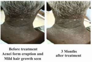
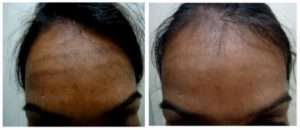
Our patients underwent anywhere from 2 to 6 treatments with the device depending on the individual clinical presentation as well as the location of the scars, until a satisfactory outcome could be achieved. (Figure 1A and 1B) It was found that regardless of the scar type, age of the scar, as well as anatomic location, fractional radiofrequency treatments could significantly improve the scar tissue in all of our patients in just a couple of sessions.

Pigmentation

Patients with stubborn melasma, post-inflammatory pigmentation as well as photomelanosis usually showed significant improvement on being treated with Fractional Radiofrequency. Diagnosis of pigmentation is quite important as it cannot be used for any malignant pigmentation, toxic pigmentation or birth marks. Once the cause of pigmentation is established, the pre-procedure preparation for the patient remained the same. For pigmentation, the parameters selected were between 220 to 245 V, with 10 to 15 msec pulse duration. The patients were then asked to apply a sunscreen and were prescribed non-steroidal depigmenting creams along with oral tranexamic acid. Some of the patients are also treated with topical vitamin C as well as topical tranexamic acid immediately after the procedure. A remarkable reduction in the pigmentation is achieved in just two sittings for most of the patients. (Figure 2a & 2B)

fractional radiofrequency

Conclusion

It was observed that majority of the patients undergoing treatment for scars as well as pigmentation showed significant improvement after a couple of sessions with fractional radiofrequency. The scars not only appeared more homogenous but also matched the surrounding non scarred skin as far as texture and colour were concerned.

While undergoing treatment, the radiofrequency energy brings about dermal heating as well as subsequent therapeutic fallout of the induced tissue inflammation. There is an increase in the temperature of the targeted skin leading to denaturation of collagen and stimulation of fibroblasts to generate new collagen and elastic fiber formation.

Amongst all the energy based devices currently available for scar and pigmentation treatment, fractional radiofrequency based devices have proven particularly successful for this indication due to their favorable aesthetic outcomes coupled with an excellent side effect profile.

References
  1. Simmons BJ, Griffith RD, Falto-Aizpurua LA, Nouri K. Use of radiofrequency in cosmetic dermatology: focus on nonablative treatment of acne scars. ClinCosmetInvestigDermatol. 2014 Dec 12;7:335-9.
  2. Lanoue J, Goldenberg G. Acne scarring: a review of cosmetic therapies. Cutis. 2015 May;95(5):276-81.
  3. Peterson JD, Palm MD, Kiripolsky MG, Guiha IC, Goldman MP. Evaluation of the effect of fractional laser with radiofrequency and fractionated radiofrequency on the improvement of acne scars.Dermatol Surg. 2011 Sep;37(9):1260-7.
  4. Min S, Park SY, Yoon JY, Suh DH. Comparison of fractional microneedling radiofrequency and bipolar radiofrequency on acne and acne scar and investigation of mechanism: comparative randomized controlled clinical trial. Arch DermatolnRes. 2015 Dec;307(10):897-904.
  5. Hongcharu W, Gold M. Expanding the clinical application of fractional radiofrequency treatment: findings on rhytides, hyperpigmentation, rosacea, and acne redness. J Drugs Dermatol. 2015 Nov;14(11):1298-304.
  6. Kaminaka C, Uede M, Matsunaka H, Furukawa F, Yamamoto Y. Clinical studies of the treatment of facial atrophic acne scars and acne with a bipolar fractional radiofrequency system. J Dermatol. 2015 Jun;42(6):580-7.
  7. Krueger N, Sadick NS. New-generation radiofrequency technology. Cutis. 2013 Jan;91(1):39-46.
  8. Kim JE, Lee HW, Kim JK, Moon SH, Ko JY, Lee MW, Chang SE. Objective evaluation of the clinical efficacy of fractional radiofrequency treatment for acne scars and enlarged pores in Asian skin. Dermatol Surg. 2014 Sep;40(9):988-95.
  9. Kaminaka C, Uede M, Makamura Y, Furukawa F, Yamamoto Y. Histological studies of facial acne and atrophic acne scars treated with a bipolar fractional radiofrequency system. J Dermatol. 2014 May;41(5):435-8.
  10. Gold MH, Biron JA. Treatment of acne scars by fractional bipolar radiofrequency energy. J Cosmet Laser Ther. 2012 Aug;14(4):172-8.

Cutis Rhomboidalis Nuche: A Rare Case

Introduction:
Abstract:

Chronic Exposure to (abstract) sunlight causes various skin manifestations and if it is a prolonged exposure as in farmers and those who take sunbath, it will end up in thickened diamond shaped skin called as cutis rhomboidalis nuche, neck being common site hence the name.

Case:

A 60 Years old male farmer came with non-itchy thickened skin over the neck and he has been suffering from the condition for the past 10 years. He gave a history of exposure to sun rays for a period of around 40 years, more than five to six hours daily.

On examination, he had hyper pigmented diamond shaped thickened skin over the neck. He came not for the cosmetic problem but he feared about the condition and worried about a malignancy. He had applied a lot of native medicine including turmeric.

Since no effective treatment is available, the patient was given the following advice:

  1. Patient consoled and given counseling.
  2. Strict advice given to avoid sun exposure.
  3. Regarding the use of fluent use of sunscreen cream once in 4 hours.
  4. He was advised to use polypodium leucomotos regularly and advised to come for review after two months.
  5. Topical retinoic cream in the night with and moisturizer.

Patient came for review 3 months later and evaluated for improvement. Moderate improvement was noted after 3 months. Photos were taken before and after treatment with the consent of the patient.

cutis rhomboidalis cutis rhomboidalis

*-*-*

Improving Feminine Health With Latest Technology

Abstract

Physiologic changes in a woman’s life, such as childbirth, weight fluctuations, and hormonal changes due to aging and menopause, may alter the laxity of the vaginal canal, damage the pelvic floor, and devitalize the mucosal tone of the vaginal wall.

These events often lead to the development of genitourinary conditions such as stress urinary incontinence, vaginal atrophy, dryness, and physiologic distress affecting a woman’s quality of life, self-confidence, and sexuality.

This review aims to present one of the available technologies “Co2 Laser Vaginal Rejuvenation” offering vaginal rejuvenation and the scientific evidence that underlines their safety and efficacy for this indication.

Introduction

STRESS URINARY INCONTINENCE is a commonly found problem in women. 35% of women over the age of 60 years suffer from Urinary Incontinence. It is caused by damage to pelvic support structures as a result of childbirth. Stress incontinence is often seen in women who have had more than one pregnancy and vaginal delivery. This results in leakage of small amount of urine with activities like coughing, sneezing, lifting, etc.

Among the new modalities being applied to feminine rejuvenation include CO2-based or erbium:yttrium-aluminum-garnet (Er:YAG) lasers and radiofrequency (RF)-based energy devices. By harnessing laser or RF waves to heat the connective tissue of the vaginal wall to 40 °C to 42 °C, these energy-based devices aim to induce collagen contraction, vascularization that ultimately revitalize and restore the elasticity and moisture of the vaginal mucosa.

Co2 Laser Vaginal Rejuvenation works via pulses of laser energy into the collagen layer of the vaginal mucosa, where concentrated thermal heating leads to collagen and elastin “shrinkage” and secondary regeneration, producing a tightening of the vaginal tissues.

How long does the result of Co2 Laser Vaginal Rejuvenation last?

Present data suggest that, with 3 treatments over a 12-week time period and especially with a “touch-up” after one year, the tightening and continence effects are experienced for at least 2-3 years or more, while the atrophic elasticity effects are ongoing with no need for further treatments in properly managed patients.

To summarize:

“Co2 Laser Vaginal Rejuvenation” is a unique technology and device which can give following advantages:

  • Proven technology for safe, accurate and effective results
  • Pain free vaginal remodeling and stress urinary incontinence
  • Lunch break procedure at the Gynecology clinic (15 – 30 min)
  • Long lasting results
  • No need for Anesthesia
Conclusion

Laser and RF technologies are widely and successfully used for treatments in dermatology and aesthetic medicine, stimulating the remodeling tissue properties and inducing the production of new collagen and elastic fibers. In the hands of well-trained physicians, energy-based devices are likely to benefit millions of women by aiding them in reclaiming, relishing, and reveling in their femininity at full capacity. 🙂

Tinea Ballads

In a faraway land, amid equatorial climes

Lies a kingdom revered from ancient times

A favorite of invaders, it was home to many a race

After battles for independence, it gradually regained its grace

This nation reborn is a mix of modern and traditional mass

With the rich and the poor and a bustling middle class

In this land, there was a dermatologist renowned

In his clinic there were always many patients to be found

Dermatology was a fantastic branch to be in

After all, the largest organ is the human skin

As the nation advanced, infections had reduced

Dr Skin was overwhelmed with work, all aesthetics induced

One day, however he saw a curious case

Rashes all over, including the face

Clear at the centre, advancing at the rim

Polycyclic margins, looked like tinea to him

He examined the patient, checked him thoroughly

Prescribed medication and sent him off early

He thought it was done, but it was not to be

For the patient returned with his entire family

Slowly but surely the scourge spread through the land

An epidemic of scratches, itches and rashes

How did it happen, he pondered and wondered

Was it because it rained heavily, clouded and thundered

Wet clothes, obese bodies and sweaty skin

That blatant steroid abuse had rendered thin

Drugs would work but for a short while,

For the tinea would be back with a vicious smile

That smile soon changed to a wide-toothed grin

For tinea mercilessly ravaged the entire skin

Allylamines, azoles or griseofulvin orally

Powders soaps and creams topically

Wash your clothes well, wash separately

Stay cool and dry, he warned desperately

But all these efforts, none seemed to work

There was always a case where tinea lurked

A nation of doers, go-getters and achievers

Was getting reduced to itchers and scratchers

The media was flooded with ads galore

Use this cream and be fairer than before

The dark side of these remedies was itchy skin

It was a losing battle, tough to win

One day, the doctor received a frantic call

The king wished to see him, the palace had called

The lord of the land, the monarch himself

Had fallen prey to tinea and was beside himself

Aha thought the doctor, this is my chance

Maybe we can end this vicious fungal dance

He examined the case, very thoroughly indeed

Did the fungal scrapings, confirmed the breed

He suggested a very effective remedy

The hapless lord was beside himself with glee

Tell me dear doc, what can I do,

You have relieved my suffering so well too

Ban offending cream ads, said the righteous man

No steroid without prescription throughout the land

This is a terrible curse, we need to fight it

Small measures won’t do, we have to right it

The wise king listened, it was a tough choice

The welfare of the people against a commercial voice

But good sense prevailed ultimately

And the doc got what he had prayed for fervently

The dreaded disease was under control at last

Dermatology was back to aesthetics, really fast

This tale was fictional but it could be true

Someday this could happen to me and you

We need to tackle the tinea bull by its horns

Or there would be no pearls in skin, just fungal thorns

*-*-*

Recalcitrant Tinea

  • Dermatophyte infections encountered in day to day practice are on the rise
  • Commonly used oral antifungal drugs fail to show response
  • True prevalence difficult to determine because of self treatment / live with chronic infection
  • Risk of acquiring such an infection is 10-20%
  • Seen in all age groups & both genders
  • Reasons for treatment failure are many
  • True prevalence difficult to determine because of self treatment / live with chronic infection
  • Poor adherence to treatment
  • Re-infection from close contacts
Host factors responsible for recalcitrant nature of infections
  • Non compliance or non adherence of schedule
  • Inadequate dosing
  • Use of tight synthetic, occlusive clothing
  • Co morbid situation – Diabetes, Obesity, Anemia
  • Immunocompromised states, atopy, thyroid
  • High risk profession – cooks and domestic help
  • Missed tinea of vellus hair on glabrous skin
  • Hidden tineaunguim

Parasitism of the hair could be the cause of the majority of isolated lesions of ringworm of hairless skin with a poor outcome with topical antifungal treatment

In some patients, some short thin hairs fell easily on to the slide during the skin scraping. On microscopy, all the few vellus hairs identified in direct examination were affected.

Excoriation may also play a role in the development of tinea of the vellus hair, probably due to scratching, making some dermatophytes located in the stratum corneum penetrate into the hair follicle.

Diagnostic clues in tinea of vellus hair
  • Inflammatory and/or excoriated lesions of tineacorporis or faciei
  • Follicular micropustules
  • Previous use of topical corticosteroids
  • Resistance to topical antifungal treatment
  • Falling of short and fine hairs to slide by scraping
Parasitism endothrix/ectothrix under potassium hydroxide examination

Dermatalk

Reference:
  • Mukherjee Pranab K. et al – Clinical Trichophytonrubrum Strain Exhibiting Primary Resistance to Terbinafine. Antimicrobial agents and Chemotherapy 2003 Jan; 47(1): 82–86
  • Diagnostic Value of selected signs and symptoms in tinea infection. AFP 1998 Jul 1;58(1): 163-174
  • Turnidge J, Paterson DL. Setting and revising antibacterial susceptibility breakpoints. ClinMicrobiol Rev. 2007;20:391-408
  • Clinical Infectious Diseases 2008; 46:120–8
  • British Journal of Dermatology 2010 163, pp603–606

A Study of Dermatoses of Pregnancy

Abstract

Background: During pregnancy profound immunologic, metabolic, endocrine and vascular changes occur resulting in both physiologic and pathologic changes in the skin and its appendages. These skin changes can be separated into three main categories namely hormone related, pre existing and pregnancy specific.

Aim: The present study was conducted with an aim to find the clinical pattern and incidence of various skin changes of pregnancy.

Methods: All pregnant females reporting to antenatal clinic of SGT Medical College and Hospital during the period of one year were included in this study.

Results: A total of 341 pregnant women were enrolled in the present one year long study out of which 194 (56.89%) were primigravidas and 147 (43.11%) were multi gravidas. Maximum incidence of 78% was found in the age group between 15-25 years of age. Physiological changes were seen in all the pregnant females while dermatoses affected by pregnancy were reported in only 12.3% cases. Dermatoses specific for pregnancy were found in 7.9% cases.

Conclusion: Clinicians must be able to distinguish physiological changes of pregnancy from several other dermatoses that can present during the period of pregnancy in order to provide timely management and thus reducing maternal and fetal morbidity.

Keywords: pregnancy, dermatoses, skin

Introduction:

Cutaneous changes and eruptions during pregnancy are exceedingly common and in some cases a cause for substantial anxiety on the part of the prospective mother. Some of these are benign and reversible after delivery whereas others can have potential effects on foetus in terms of morbidity and mortality.[1] Likewise, The concerns of the patient may range from cosmetic appearance, to chance of recurrence of the particular problem during a subsequent pregnancy, to its potential risks on fetus.[2] There are three general categories of pregnancy associated skin conditions: 1) benign skin conditions from normal hormonal changes or physiological skin changes, 2) pre existing skin conditions that change during pregnancy or skin diseases affected by pregnancy, and 3) pregnancy specific dermatoses.[3] Physiological skin changes include striae gravidarum, hyper pigmentation and hair, nail and vascular changes. Pre existing skin conditions that may be affected by pregnancy include psoriasis, atopic dermatitis, candidal and other fungal infections, benign appendageal lesions and many more. True dermatoses of pregnancy include pruritic urticarial apples and plaques of pregnancy (PUPPP), prurigo of pregnancy, intrahepaticcholestasis of pregnancy, pemphigus gestationis, impetigo herpetiformis and pruritic folliculitis of pregnancy.

Materials and Methods:

The study comprised all pregnant females reporting to antenatal clinic of SGT Medical College and Hospital during the period between January 2015 to December 2015 were included in this study. A total of 346 pregnant women were included in the study. Informed consent was obtained before before the interview and clinical examination.

All patients were subjected to detailed history and thorough clinical examination to note all the physiological and pathological mucocutaneous changes. Routine blood, urine and stool examination, and screening with VDRL and ELISA for HIV was done in all cases. In patients with white discharge, a KOH mount, saline mount and gram stain were done.

Results:

A total of 341 pregnant females were included in the one year study conducted between January 2015 to December 2015. Patients were divided in age groups between 15-25 years (266 cases, 78%) and 26-40 years (75 cases, 22%). Of these, 194 (56.89%) females were primigravidas and 147 (4.11%) were multi gravidas. Most of them presented in their third trimester (222, 65.1%) followed by second (75, 22%) and first trimester (44, 12.9%).

Pruritus was the commonest symptom reported accounting for 18.2% (62) of the patients, followed by white discharge per vagina (21, 6.1%), while few complained of presence of one or other type of skin lesions due to dermatoses which were broadly divided into three categories:

Physiological cutaneous changes Skin diseases (Pre existing or newly occurring) affected by pregnancy.

Pregnancy specific dermatological disorders.

Physiological changes were seen in 100% of cases, while dermatoses modified by pregnancy were observed in 12.3% cases and only 7.9% had dermatoses specific for pregnancy.

Among the physiological changes, most common were the pigmentary changes including lineanigra, areolar pigmentation and melasma followed by connective tissue changes mainly including striae gravidarum.[Table 1]

Candidal vaginitis (13, 3.81%) was found to be the most common dermatosis affected by pregnancy closely followed by acne vulgaris (9, 2.64%) and eczemas (6, 1.76%).[Table 2]

Total of 37 females were diagnosed of pregnancy specific dermatoses of which 19 (5.57%) had PUPPP (pruritic urticarial papules and plaques of pregnancy).[Table3]


Discussion:

Pruritus was reported to be the commonest symptom with incidence of 18.2% in the present study. According to Winton et al, pruritus from all cases may occur in 17% of pregnant women,[4] while Wong et al and Roger et al reported incidence of pruritus in 20% and 18% respectively.[5,6]

Candidal vaginitis was found to be the most common cause for vaginal discharge. That monilial vaginitis is 10 to 20 times more common during pregnancy has been supported by Winton et al and Dotz et al.[1,7]

Physiological changes were seen in all the pregnant females. Several other studies have reported physiological changes in upto 100% cases.[8,9] Pigmentary changes were noticed in all the cases.[3] Striae were found to be the commonest physiological change (87.68%) closely followed by Lineanigra (85.3%) and areolar hyper pigmentation (82.1%) [Fig 2]. Other less commonly encountered physiological changes were melasma, gingivitis, acne, montgomery tubercles, hair and nail changes.

The most common physiological changes are pigmentary alterations, stretch marks, vascular spiders and telogen effluvium.[10] Pigmentary changes occurred in 98.82% of the patients.[11] Similar incidences have been reported by other authors.[4,5,7] Incidence of melasma was reported to be 10% similar to the finding of V. Shivakumar and Raj et al.[11,12]

In our study, striae gravidarum were seen in 299 (87.68%) cases. A few studies have shown incidences between 60 to 80% [8,9,11,12] while others have reported incidences upto 90%.[1,5,7]

Montgomery’s tubercles is well known during pregnancy in 30-50% of pregnant women. [10] In our study, Montgomery’s tubercles were seen in 22 (6.45%) cases. This was found to be consistent with other studies. [13]

Vascular changes result from distension, instability and proliferation of vessels [4] and were seen in 45.16 % of cases. Non pitting edema over the hands, feet and face was observed in 35.78% cases. Muzaffer et al [8] reported edema in 48.5% whereas Rashmi et al [9] reported it in 9.8%. Varicosities were seen in 1.17% comparable to other studies. [8,12]

Gingivitis may occur in upto 100% of pregnant women with varying degree of severity.[5] 6.15% cases of gingivitis were reported. A bit higher percentage has been reported by other authors.[8,13]

PUPPP (also known as polymorphic eruption of pregnancy) was the commonest specific disorder of pregnancy accounting for 5.57% followed by prurigo of pregnancy with 12 (3.52%) cases and pruritus gravidarum (also known as intrahepaticcholestasis of pregnancy) with 4 (1.17%) cases. Similar to our study, PUPPP was reported as commonest specific dermatoses of pregnancy in study by Kumari R [9] accounting for 63.6% cases followed by pruritus gravidarum while Shivakumar and Madhavamurthy [11] reported prurigo of pregnancy as the commonest specific dermatoses of pregnancy with incidence of 9.41% followed by pruritus gravidarum.

Conclusion:

To conclude, a variety of cutaneous lesions can be seen in pregnant women which need a meticulous examination and investigations to reach a correct diagnosis enabling careful management in an effort to minimise maternal and foetal morbidity.

References:
  1. Kroumpouzos G, Cohen LM. Dermatoses of pregnancy. J Am Acad Dermatol 2001;45:1-19. [PUBMED]  [FULLTEXT]
  2. Lawley TJ, Yancey KB. Skin changes and diseases in pregnancy. In: Freedberg IM, Eisen AZ, Wolff K, Austen KF, Goldsmith LA, Katz SI, et al , editors. Fitzpatrick’s Dermatology in general medicine. 5th ed. McGraw-Hill: New York; 1999. p. 1963 9.
  3. Marc Tunzi, Gary R. Gray. Am Fam Physician. 2007 Jan 15;75(2):211-218.
  4. Winton GB, Lewis CW. Dermatoses of pregnancy. J Am Acad Dermatol 1982;6:977 998.  [PUBMED]  [FULLTEXT]
  5. Wong RC, Ellis CN. Physiologic skin changes in pregnancy. J Am Acad Dermatol 1984;10:929-940.  [PUBMED]  [FULLTEXT]
  6. Roger D, Vaillant L, Fognon A, et al. Specific pruritic diseases of pregnancy. Arch Dermatol 1994;130:734-739.
  7. Dotz W, Berman B. Dermatologic problems of pregnancy. In: Cherry SH, Merkatz IR, editors. Complications of Pregnancy: Medical Surgical Gynaecologic Psychosocial and Perinatal, 4th ed. Baltimore : Williams and Wilkins, 1991:562 587.  Back to cited text no. 6
  8. Muzaffar F, Hussain I, Haroon TS. Physiologic skin changes during pregnancy: A study of 140 cases. Int J Dermatol 1998;37:429-31.[PUBMED]
  9. Kumari R, Jaisankar TJ, Thappa DM. A clinical study of skin changes in pregnancy. Indian J Dermatol VenereolLeprol 2007;73:141.
  10. Martin AG, Leal-Khouri S. Physiological skin changes associated with pregnancy. Int J Dermatol 1992;31:375-8.
  11. Shivakumar V, Madhavamurthy P. Skin in pregnancy. Indian J Dermatol VenereolLeprol 1999;65:23-5.
  12. Raj S, Khopkar V, Kapasi A, et al. Skin in pregnancy. Indian J Dermatol VenereolLeprol 1992;58:84-88.
  13. Rathore SP, Gupta S, Gupta V. Pattern and prevalence of physiological cutaneous changes in pregnancy: A study of 2000 antenatal women. Indian J Dermatol VenereolLeprol 2011;77:402.

Dermatoses

* more than one dermatoses were present in many cases

Table 1: physiological changes of pregnancy

Dermatoses

Table 2: Dermatoses affected by pregnancy

Dermatoses

Table 3: pregnancy specific dermatoses

*-*-*