Childhood Vitiligo

Vitiligo is a skin condition characterized by patches of skin losing their pigmentation, due to the destruction of the pigment producing melanocytes. This disease affects an estimated 1-3% of the world’s population, and has a strong familial association. Half the patients first recognize Vitiligo before 20 years of age, when it often appears in an area of minor injury or sunburn.

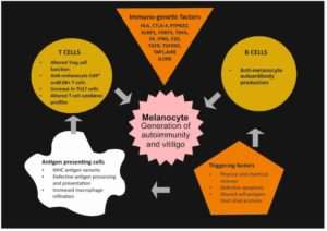
The central process in Vitiligo is the destruction of melanocytes at the dermoepidermal junction. Fully blossomed lesions are totally devoid of melanocytes. Rarely, a superficial perivascular lichenoid mononuclear infiltration may be observed at the border of the depigmented areas. The exact cause of Vitiligo is unknown. Autoimmune mechanisms with an underlying genetic predisposition are the most likely cause. Antibodies to the melanocytes have been detected by immunoprecipitation in the sera of the patients with Vitiligo. Also, sera from patients with Vitiligo causes damage to melanocytes in cell culture, suggesting that the antibodies present in sera of affected patients are involved in the pathogenesis of Vitiligo.

Childhood Vitiligo

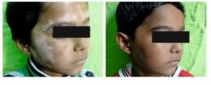
Childhood Vitiligo differs from adult-onset Vitiligo for several features including increased incidence of the segmental variant, higher prevalence of halo nevi, and more common family history for autoimmune diseases and atopic diathesis. Trigeminal area is the most common site involved. Half the cases are associated with white hair (poliosis).

Types of Vitiligo

The most characteristic types include generalized vitiligo, focal vitiligo, acral vitiligo, acrofacial vitiligo, segmental vitiligo and vitiligo universalis. The segmental type of vitiligo is more often seen in children, as it starts generally earlier in life than the non-segmental one.

Associated Comorbidities in Childhood Vitiligo

Other cutaneous abnormalities include leukotrichia, premature gray hair, halo naevi and alopecia areata. Iris and retinal pigmentary abnormalities and deafness may be present. Systemic diseases associated include thyroid disease, diabetes mellitus, pernicious anemia, Addison’s disease and multiple endocrinopathy syndrome. Presence of leukotrichia, associated with autoimmune disorders and family history are poor prognostic factors. Hashimoto’s thyroiditis is 2.5 times more frequent among children and adolescents with Vitiligo than in a healthy age and sex-matched population. It usually follows the onset of Vitiligo. A family history of vitiligo is associated with earlier age of onset, among pediatric patients, those with a positive family history are more likely to develop Vitiligo before 7 years of age.

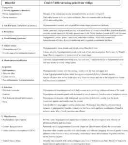
The major differential diagnoses are the post inflammatory hypomelanoses for nonsegmental Vitiligo and nevus depigmentosus for segmental Vitiligo.

Pathogenesis of Vitiligo

childhood vitiligo 2

Clinical features of Vitiligo

The most common presentation of Vitiligo is totally amelenotic (i.e. milk or chalk white) macules or patches surrounded by normal skin. The lesions characteristically have discrete margins, and they can be round, oval, irregular or linear in shape. The borders are usually convex, if the depigmenting process was ‘invading’ the surrounding normally pigmented skin. The lesions enlarge centrifugally over time, and the rate may be slow or rapid. Vitiligo macules and patches range from millimeters to centimeters in diameter and often vary in size within areas of involvement. The incidence of leukotrichia varies from 10% to more than 60%, as follicular elanocytes are often spared in vitiligo.

Any of the clinical variants of vitiligo may occur in childhood. Vitiligo vulgaris is the most common clinical type observed in various clinical studies, followed by focal vitiligo and segmental vitiligo. Acrofacial and mucosal vitiligo have a lower incidence in childhood. The rarest type seen during childhood is the universal vitiligo.

Common initial site of onset of both NSV and SV in children is the face and neck. In NSV, initial lesions are periocular, perinasal or perioral, and gradually spread to other body parts, in a more or less symmetrical manner. Perineum, perianal area and, in infancy, the diaper area may be the initial site of occurrence of skin lesions. Individual vitiligo macules may enlarge attaining a geographic pattern or there may be appearance of new lesions at other sites. Although extensive areas of depigmentation may be present, majority of the children have <20% body surface area (BSA) involvement. Focal vitiligo may subsequently evolve into generalized disease.

Koebner phenomenon may be noted in both SV and NSV in children, more commonly in the latter type. In SV, Koebnerization is confined only to the involved segment. Koebnerization may be more frequent in childhood vitiligo because of higher mobility and playfulness in this age group. Presence of Koebnerization is indicative of the disease activity.

childhood vitiligo 3

Diagnosis of Vitiligo

Diagnosis of Vitiligo is mostly clinical. In children with fair skin, it may be difficult to differentiate a lesion of Vitiligo from the surrounding normal skin. In these cases, examination under Wood’s lamp is helpful. Complete blood count and fasting blood sugar should be performed as a routine work-up for all patients. In case of diagnostic difficulty, skin biopsy may be taken; histopathological examination shows total absence of melanocytes in established lesions of Vitiligo.

In early lesions, melanocytes are still retained but with multiple abnormalities like vacuolization, dilated endoplasmic reticulum and granular deposits. Presence of mild inflammatory infiltrate is sometimes seen, and is indicative of disease activity.

Associated autoimmune disorders should be ruled out in the pediatric age group. Screening for autoantibodies may be performed if facilities are available. Antinuclear antibody may be positive even in normal children. Thyroid function status of the child is assessed by estimating T3, T4 and TSH levels. Coexistence of autoimmune thyroiditis may be ruled out by the estimation of antithyroglobulin antibody (anti-Tg) and antithyroperoxidase antibody (anti-TPO).

Differential Diagnosis of Childhood Vitiligo

Childhood vitiligo-dermatalk

Vitiligo and Quality of Life in Children

Study shows that vitiligo is related to depressive symptoms in children. A similar effect was not detected in adolescents, even though Vitiligo has a disfiguring and stigmatizing nature, which could affect identity formation and self-definition.

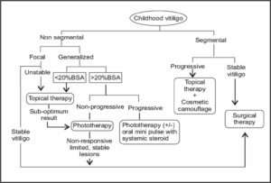
Treatment of Childhood Vitiligo

Various therapeutic modalities are available for the treatment of Vitiligo. However, all of these cannot be used with respect to children. Medical therapy is considered as the first line of management in this age group. All children with widespread Vitiligo should undertake photoprotection preferably with opaque sunscreens during daytime outdoor activities. In general, localized Vitiligo is treated with topical therapy. Widespread or generalized disease is managed with phototherapy or systemic therapy. Surgical methods may be chosen for stable (size of the lesion is stationary for > 2 years and no new lesion has developed recently) localized Vitiligo and SV. Certain clinical features are poor prognostic markers for treatment. These include acral Vitiligo, presence of leukotrichia over vitiliginous area and lesions over bony prominences like elbow, knee and ankle.

Following table shows various therapy for Vitiligo
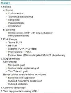
vitiligo

Children vitiligo-dermatalk

Medical Treatment

Topical Therapy

Mid-potent topical corticosteroids are the first-line therapy for children with localized Vitiligo. Although high-potency steroids are more effective in Vitiligo, these are not recommended for use in children. It requires long-term therapy for several months, increasing the chances of developing tachyphylaxis and local side effects like atrophy, telangiectasia, hypertrichosis and striae. There are risks of serious side effects like glaucoma (prolonged application on periorbital Vitiligo), suppression of hypothalamic-pituitary-adrenal axis and growth retardation (longterm use over large BSA). Hence, parents should be cautioned about the inadvertent use of topical steroids.

Topical calcineurin inhibitors (TCI), tacrolimus and pimecrolimus are effective alternatives to topical corticosteroids in terms of avoidance of side effects of the latter.

In an open comparative trial of mometasone cream (0.1%, once-daily application) and pimecrolimus cream (1%, twice-daily application) in the treatment of localized childhood Vitiligo, the repigmentation rates were 65% and 42%, respectively, at the end of 3 months. Mometasone cream was found to be equally effective in all body parts whereas pimecrolimus was effective only over the face.

TCIs are slower to exert beneficial effect compared to topical corticosteroid. Best response is observed on the thinnest areas of the skin (eyelids). As the skin barrier function is not compromised in children with vitiligo (unlike atopic dermatitis), highest strength (0.1%) of tacrolimus may be used safely. Calcineurin inhibitors are not yet approved for the treatment of Vitiligo and are not recommended for use in children <2 years.

Topical calcipotriol has been found to be effective in the treatment of Vitiligo in children and adolescents. In a trial of topical calcipotriol in childhood Vitiligo, repigmentation was recorded as early as 4 weeks of treatment. Combination therapy of topical corticosteroids and calcipotriol may be used to reduce the side effects of corticosteroids as well as to enhance the efficacy of calcipotriol as the latter may not be as effective when used as monotherapy. Side effects of calcineurin inhibitors and calcipotriol are mild and transient and
hence, easily tolerated by children. Recalcitrant cases of widespread Vitiligo or universal Vitiligo with only a few islands of normal skin may be considered for total depigmentation therapy with 20% monobenzyl ether of hydroquinone (MBEH), with an advice of life-long stringent photoprotection.

Systemic Therapy

Rapidly progressive generalized Vitiligo in older children and adolescents may be treated with a short course of systemic steroid. A better way to avert the side effects is administering oral betamethasone as a single morning dose (0.1 mg/kg body weight) on two consecutive days in a week (oral mini-pulse therapy) for 12 weeks, and thereafter reducing the dosage by 1 mg/month for the next 3 months. Pasricha et al have studied the effect of oral mini-pulse therapy (OMP) in children and adults with extensive or fast-spreading Vitiligo, and have observed a 26-50% response in 25%, 51-75% response in 7.5% and >75% response in 15% of patients. Phototherapy may be added to such regimen. Majid et al have used methylprednisolone OMP in combination with topical fluticasone for 6 months in 400 children with progressive vitiligo. Complete halt of progression was noted in >90% of the children after initiation of therapy, and >65% of children had well to excellent repigmentation at the end of the study period.

Phototherapy and Photochemotherapy

Systemic photochemotherapy with psoralen-ultraviolet A (PUVA) is contraindicated in small children and can be used only beyond 12 years of age. It is used when >20-25% of the BSA is involved or in patients in whom other treatment modalities are not giving optimum result. Topical PUVA can be used even in younger children with limited BSA involvement. Various authors have reported an overall response rate of approximately 75% in 50-60% of the children with Vitiligo treated with topical or oral PUVA therapy.

Oral supplementation of L-phenylalanine (a precursor of tyrosine in melanin synthesis) and UVA phototherapy have been found to be effective in children with extensive Vitiligo (50-100% repigmentation in 69% of children).

Narrow-band ultraviolet B (NB-UVB) phototherapy is an effective modality for the treatment of generalized childhood Vitiligo with >20% BSA involvement. There are several studies on the effectiveness of NB-UVB phototherapy in children. Brazzelli et al have reported this therapeutic modality as effective and safe in children. Best repigmentation occurred on skin lesions over the face and neck, and moderate response was seen in lesions on the trunk and proximal extremities. Lesions over the acral region (fingers and toes), bony prominences and less-hairy areas (wrist, ankle and joints) were poorly repigmented. The side effects recorded were minimal erythema, amenable to emollients and topical steroid.

Excimer laser (308 nm)/targeted UVB phototherapy has been used in the treatment of localized childhood Vitiligo with the advantage of more focused therapy at the site of lesion, avoiding the risk of photoageing of the surrounding skin. Cho et al have treated 30 children with localized Vitiligo (40 patches) with 308 nm excimer laser. A repigmentation rate of >50-75% was recorded in this trial, especially over the face, neck and trunk. Side effects related to this mode of therapy are usually minimal and transient. Repigmentation following phototherapy is either marginal or perifollicular.

During phototherapy, genitalia of the children should always be protected. It is preferable to follow the “skin-saving principle,” i.e. shielding of uninvolved body sites during phototherapy. Already repigmented body parts should also be covered with clothing during further therapy. Carcinogenic potential of NB-UVB phototherapy in children is not yet determined. However, theoretically, there may be an increased chance of developing long-term skin cancers because of the higher number of expected years of life in children. Such risk may be lower in children with darker skin types. In resource-poor countries, the facility of phototherapy is available mostly in tertiary health care centers. Hence, it may not be easily accessible to families from remote localities. Moreover, multiple hospital visits confer loss of school days of children and working days of parents, reducing compliance to treatment.

Surgical Treatment

Only stable localized Vitiligo lesions (segmental or non-segmental), unresponsive to other treatment modalities, are chosen for surgical treatment. Surgical procedures are not performed in very young children. This is because segmental or stable focal lesions in younger children extend proportionate to their body growth. Moreover, success of many surgical procedures depends upon the postoperative immobility of the operated part, which becomes difficult to maintain in young children. Older children and adolescents may be counseled about the procedure and the possible outcome of the surgery to achieve their cooperation. The restrictive factors for surgical procedures are inability to treat larger area and the risk of Koebnerization of the donor site.

Among the various surgical techniques, suction blister epidermal grafting (SBEG) has been found to be most convenient and effective for children and adolescents. The success rate of this technique is better in children as compared to adults. However, this procedure requires prolonged immobility to facilitate smooth generation of the blisters, more so in children because of their strong dermoepidermal adherence. Hence, it may be a difficult task to keep children in restricted posture till the required duration. Surgical procedures in combination with medical therapy have been tried in childhood vitiligo. Non-cultured autologous epidermal transplantation has been used successfully in children and adolescents with stable vitiligo. Cultured melanocyte transplantation is a relatively tedious technique requiring specialized set up, trained staff and a preparation time of 6-8 weeks.

Cosmetic Camouflage

Good-quality cover-up cosmetics (Dermacolor) may be used to cover localized Vitiligo lesions over exposed body parts in children. Well-counseled older children may use this as an effective adjunctive while on treatment or when failed to respond to other treatment modalities.

What’s New in Vitiligo?

Latanoprost, commonly used in the treatment of ocular hypertension was tried in Vitiligo based on the presence of irreversible iris pigmentation and reversible periocular hyperpigmentation observed in these patients. PGF2α, however, exerts its effect indirectly through induction of COX-2 and PGE2 and has been reported to be a promising therapeutic option for Vitiligo in both animal and human studies with increased efficacy when combined with phototherapy. Other Treatment Methotrexate, known for its use in inflammatory and immunemediated conditions, such as psoriasis and inflammatory bowel disease, has also been studied in treating Vitiligo. In childhood Vitiligo, it should be used with care.

Tofacitinib is a recently approved drug for rheumatoid arthritis and acts as a Jak inhibitor, disrupting specific inflammatory pathways that signal through proteins called Jaks, of which there are four – Jak1, Jak2, Jak3, and Tyk2. While only a single patient, this report is indeed very exciting, as it suggests that Jak inhibitors may provide a new effective treatment for Vitiligo. IFN-g signaling pathway (which requires Jak1 and Jak2) is critical for vitiligo progression and maintenance.

Conclusion

Over the last decade, significant progress has been made in understanding the complex and multifactorial pathogenesis of Vitiligo. It is now theorized that intrinsic metabolic, neuronal, and/or biochemical cutaneous perturbations trigger autoimmune melanocytic destruction, to which patients may be predisposed by certain genetic mutations and polymorphisms. How different pathogenic mechanisms give rise to Vitiligo subtypes remains to be further elucidated. There is no cure for vitiligo at present. However, a variety of advances in medical and surgical treatments as well as combinations of the two working synergistically have shown the ability to improve patients’ disease state and quality of life. A number of clinical trials are underway, and the future looks promising for patients afflicted with this traditionally stigmatizing and challenging condition.

World Vitiligo Theme 2017

Step up for Vitiligo: A Call for Truth, Hope and Change!

Complication And Management In Hair Transplant

ABSTRACT: 

Surgical complications in hair transplant are at times a serious matter. After a follow up for one year post operation, following were the complications: Post operation edema, puriritis, numbness, paraesthesia, bleeding, wide donor scar, sterile folliculitis. Good communication between patient and surgeon, complete clinical and lab assessment of patient, accurate surgical technique trained surgical team are crucial for successful hair transplant with minimal complication rate.

Definition: 

Complication is defined as an adverse event that is not considered to be common and which requires change in methodology.

It may be due to surgeon’s planning and technical error or because of patient’s physiology or compliance error.

Surgical Technique: 

After ring block in donor area under aseptic condition in patient with stable medical condition.

FUT: 

A strip of skin along with hair is cut from the donor area and then the hair is cut into grafts and then the gap in the donor area is stitched by Thchophytic closures.

hair transplant

After the graft is extracted it is implanted in the recipient area with the help of needle and forceps.

FUE: 

Extraction is done from the safe Donor area with the help of motorised blunt punches or sharp Titanium manual punches.

hair transplant 2

After the extraction the graft is implanted in the recipient area with the help of needles and forceps or implanter.

Complications and Discussion:

1. Post Operation Oedema: (most common complication)

This occurs on third day of surgery and use of steroid in tapering doses decreases the swelling and inflammation.

Limited use of tumescent anesthesia reduces the chance of post operation oedema. Addition of 1ml adrenaline (1:1000), 2ml Xylocaine -2%, 1ml triamcilone (40mg/ml) in 100 ml. Saline for tumescent anaesthesia

Swelling occurs on the forehead then around eyes and then on the face and is absorbed in a week or 4 to 7 days.

hair transplant 3

2. Bleeding:(second most common complication)

Stop anabolic steroids, vitamin E, Ginko biloba, Aspirin 10 day prior. NSAID and alcohol – a day prior to decrease risk of bruising and oedema. Vitamin – C per week 2000mg OD BT, CT, PT should be normal and patient should be on Warfarin or Heparin injection.

3. Pruritis:(third most common complication)

This occurs up till 12 week post operation which can be controlled by Antihistamaine as and when required.

4.Numbness Paraesthesia: 

Hypoaesthesia, Neuralgia is due to the nerve damage, most commonly in FUT but sometimes Neuralgia is common in diabetic patients undergoing FUE up to 3 – 18 weeks.

5. Wide Donor Scar:

This is common in FUT when the strip is wide and too much of tension is there during closer l/t Hypertrophic scar, keloid.

Mayers Paul scalp, elasticity scale measures the laxity of donors scalp. This scale is guide for estimation of Donor area that can be safely closed with minimum tension.

hair transplant 4

If a wound closure is in tension, it can lead to necrosis, unpredictable hair loss in donor area and scarring.

In FUE punches ≤ 1 mm should be used and better to punch 1 in every 5th hair and try to cover the entire area even though the number of graft to be extracted is less to avoid moth eaten appearance in the donor area.

6. Sterile Folliculitis: The common cause is in-growing of hair, foreign body reaction epithelium logged into stilt site or piggy backed graft. This is managed by careful insertion of grafts, antibiotics and unrooting of cyst. Sometimes pyogenic granuloma is seen, in such cases. Deroofing of the cyst is a must for complete cure.

Clean and decontaminated operating room, use of sterilized materials, donor area asepsis, disposable instruments, Antibiotics prophylax are vital to prevent infection.

Proper post operation care from patient side is also important for a week or two.

7. Poor Hair Growth: It depends upon the technique of graft desication in FUT.

Rough handling of graft,  prolonged out of body time, harmful effects of sterile water, folliculitis, dense placement of graft more than 40-50grafts/sq.cm. Ideal graft placement is 15-25grafts/sq.cm.

Fall of existing hair –DHT created when six reduction converts testosterone to DHT which takes place in blood and locally in the scalp. This DHT acts on genetically susceptible hair follicle to cause miniaturization of hand follicular death.

Koebner phenomenon- d/t surgery trauma. Hair follicle antigen liberated during surgery or post surgery induces hair follicles immune cascade l/t. follicular damage.

Diseased recipient area destroys transplanted hair follicles, so in active Lichen planopilaris, DLE, transplant should be avoided.

8. Cosmetically Unacceptable Result:

Use of large graft and unaesthetic hairline design, failure to anticipate future hair loss in planning, hair line restoration result in unnatural appearance as the patient age increases (large bald areas between lateral aspect of parietal hairline and temporal peak caused by recession of temporal areas).

hair transplant 5

Transplant in crown areas in young individuals result in circular region of transplanted hair surrounded by rim of baldness. When baldness increases as the age advances.

This can be corrected by placement of 1 and 2 hair graft placed in proper direction to create an irregular hairline of gradual increasing density in the center and insert a few grafts beyond the border of the existing hair, so that merging of the hair is done.

Conclusion:

Successful hair transplantation and decreased rate of complication depends on good communication between surgeon and  patient, careful clinical and lab evaluation, carefully selected technique, trained surgical team and rigorous post operation evaluation.

References
  1. Konior RJ. Complication in hair restoration surgery. Facial Plast Surg Clin North Am. 2013;21:505-20 [PubMed]
  2. David PM. Complication in hair restoration surgery. Oral Maxillofac Surg Clin North Am. 2009;21:119-48 [PubMed]
  3. Musgrave R Gradinger G Symposium on Problems and complications in Aesthetic QPlastic Surgery of the faceSt. Lois: Mosby; 1984.pp.3-5
  4. Lam SM.. Complication in hair restoration surgery. Facial Plast Surg Clin zNorth Am. 2013;21:675-80. [ PubMed]
  5. Coleman WP.3rd.Kllein JA. Use of the tumescent technique for  scalp  surgery, dermabrasion and soft tissue reconstruction. J Dermatol Surg Oncol. 1992;18;130-5.-[ PubMed]
  6. Unger WP, 3rd ed. Wew York: M Dekkaer:1997

STRIP Versus FUE Consideration

We have two excellent methods of harvesting donor hair follicles in hair restoration surgery. With follicular unit transplantation (FUT), more correctly described as the strip harvesting method, a narrow piece of scalp (typically 1cm or wider) is excised from the donor region. Surgical assistants dissect the strip with stereo microscope and result is the production of grafts that contain follicular group of 1-4 hairs. With FUE, the donor grafts are harvested individually with very small circular punches typically < 1 mm in diameter. When harvesting grafts using the FUE method, the physician creates all the grafts each containing follicular groups of 1-4 hairs.

Comparison of Techniques

So follicular unit extraction(FUE) is best choice in following cases-

  1. A patient wants to have least painful post-operative course.
  2. The scalp is very inelastic.
  3. A patient wants to start gym as early as possible.
  4. A patient wants to keep hair short (buzz cut) which is very popular nowadays, and wants to avoid linear scar.
  5. A patient has donor healing problem from previous strip surgery.
  6. Body hair needs to be harvested.
  7. Finer hair needs to be selected for hairline or eyebrow.
  8. A patient has a history of keloid or hypertrophic scarring.
  9. Repairing prior strip scar.
  10. A patient has low donor density.

Strip is best choice in following cases:

  1. A large treatment is required.
  2. A patient who wants most hair possible single day procedure.
  3. A patient has no plan or desire to keep the hair short and is unconcerned about a linear scar.
  4. A patient who does not want to shave donor region for the procedure.
  5. Cost is a major concern.

Finally it is important to note that strip and FUE are complementary techniques. They can be interchanged sequentially over a treatment course depending on the specific requirements of the patient at any given point in time. They can also be used simultaneously in same procedure to facilitate a large harvest. A hair transplant surgeon must know both the techniques.

Trichology: An Overview

Today’s world is inching towards more of a cosmetic era. Hair is considered a major aesthetic display feature of human appearance. Today hair loss or thinning is a common complaint in clinical practice.

The impact of hair loss on a patient confidence varies and they seek medical advice most frequently nowadays as there are profound psychological effects, poor self image and loss of self confidence.  In trichology practice, androgenic alopecia, telogen effluvium, female pattern baldness, seborrhoeic dermatitis and Alopecia areata are most common problems presented to a dermatologist.

Here, in this article, I have tried to give an overview, a systematic approach to history taking, rough diagnostic test and few salient treatment approach points.

History Taking:

A detailed history includes the chief complaints, past medical/surgical history, addictions, allergies, medications, diet, assessment of stress and family history. Menstrual history, pregnancy and menopause should be assessed in all female patients.

Proper history taking leads to diagnosis of cause. Most common causes are as follows:

  • Family history – male or female pattern baldness
  • Stress including emotional distress, anxiety, physical stress from surgery, illness, high fever, postpartum
  • Chemotherapy
  • Seborrheic dermatitis of scalp
  • Poor diet esp. iron and proteins deficiency
  • Thyroid diseases such as hypothyroidism and hyperthyroidism, LE
  • Tinea capitis/Psoriatic scalp/Alopecia Areata
  • Hair care practices, use of hair cosmetics, bleaching or other cosmetic procedures) hair styling, frizzy hair, few causatives of hair
  • Damage by traction due to fight braiding and clips or using curling irons, dyes

Approximate number of hair lost per day more than 100 (50-100 strand loss/day is normal) or visible scarcity or decrease in hair density need to treated.

Clinical Examination:

For global appearance, texture, color, length, it’s performed in 3 stages.

Stage I: examination of scalp for inflammation, scaling, erythema and follicular opening scarring Alopecia are devoid for follicular units.

Stage II: Distribution pattern and density of hair, focal hair loss in Alopecia Areata. Diffuse hair loss, with scalp hair distribution by scoring system like Hamilton Norwood and Ludwig classification.

Stage III: study the quality of hair shaft (caliber, fragility, length and shape). Tapered hair indicates new growth. Miniaturized hair is also tapered at end but much finer in caliber.

Hair assessment is understood by:

  1. Hair growth assessment
  2. Structural and functional assessment
  3. Scalp biopsy
  4. LAB test
A. Hair growth assessment done by:
  1. Pull Test: Bundle of 50-60 strands grasped between thumb, index & middle fingers from near the base pulled non forcibly, if 10% are pulled away it suggests positive pull test and implies active hair shedding, if less then normal physiological shedding.
  2. Modified Wash test: After 5 days’ abstention from shampooing, the patient washes and lost hair is collected and analyzed. I) Long Hair (> 5cm) 2) intermediate (> 3 to 5 Cm) 3) Short vellus hair (<3 cm). Hair shorter than 3 cm is telogen vellus hair. Suggestive of four possible diagnosis AGA, TE, AGA + TE and remittent TE.
  3. Global Photography
  4. Unit area trichogram 
  5. Trichoscopy

trichology

trichology 1

trichology

B. Structural and Functional:

Assessment is done by optical light and polarized microscopy and electron microscopy. For hair shaft defects like tapered end, broken hair, pili torti.

C. Scalp Biopsy:

It is an important tool in hair analysis in non-scarring alopecia a punch in the center of the lesion suitable whereas in scarring alopecia sample from the active peripheral margin taken.

D. Laboratory Tests:

It is carried out to support diagnosis or to plan treatment. Basic evolution for

  • Thyroid dysfunction – Thyroid profile
  • Iron deficiency anaemia – Serum iron and ferritin
  • In women with andnogenetic alopeina and virilising signs such as acne, hirsutism and / or irregular menses. Endocrine panel – free testosterone, prolactin, 17 hydroxy progesterone, cortisol to R/o hyper androgenic states.
  • For scarring alopecia secondary to DLE and LE – antinuclear antibody analysis done
  • For syphilitic alopecia – a rapid reagin test done

Therapeutic Armamentanium in Androgenic Alopecia

  • Topical – Minoxidil
  • Peptides
  • Retinoids
  • Systemic – Finasteride / Dutastende
  • Cyclical Nutrient Therapy
  • Stimulants – Laser helmet
  • PRP mesotherapy
  • Procedural – Hair Transplantation (FUE/FUT/FUSS)

(Patient should have sufficient donor area in the occipital area}

  • Scalp Reduction / resection
  • Scalp Hygiene – Anti dandruff shampoo and daily use of shampoo

A few salient points helpful in treatment are that minoxidil is still the gold standard in treatment of AGA by causing vasodilation starting with higher concentration 10% / 12.5 %, following therapeutic results slowly tapering the concentration to 7.5 and the 5% in male and 2 % in females preferably using high concentration for once at bed time eg 10% in males and 5% in females.

If patient is allergic to alcohol based minoxidil lotion, you may use gel based product.

Minoxidil with tretinoin also found quite superior in practical therapeutic effect.

Topical finasteride has no added advantage but in fact may increase the side effects if given along with oral finasteride.

  • Saw Palmetto, biotin, tripeptides, tetrapeplides, caffeine, cococin and herbs may be improving hair anchoring, stimulating fibroblast, inhibiting 5αreductase, and improving absorption of minoxidil.
  • Cyclical therapy of micro nutrients is used to deal with hidden hunger. On a daily basis, medication with B Complex, biotin, amino acids and antioxidants, while calcium, and iron on alternate day will achieve their maximum absorption and folic acid – twice a week. This helps in conversion of telogen phase to anagen phase by rapid cell division and prolonging Anagen phase. Biotin and B complex help catalyse the metabolic process.

Laser Helmet:

Hair growth with special low light laser therapy (LLLT) is US FDA approved for treatment for hair loss.

  • Acts by improving the blood flow and oxygenation to hair roots.
  • Controls over activity of sebum glands, reduces irritation, inflammation and reduces dandruff.
  • More than 30% of hair which are dormant or in catagen phase are stimulated and saved from becoming dead.

Mesotherapy by PRP:

Platelet concentration is 10 lacs / cubic mm in 5 ml PRP. There is a minimum 4 fold increase in baseline platelet concentration. It helps in stimulation by growth factors.

It is done by point by point injection technique or using dermoroller before or after application of PRP on the scalp. It requires a monthly sitting for first 3 months and later after 45 days to 2 months. Total 6 to 8 sittings recommended  depending on grade of Androgenic alopecia, female pattern alopecia and telogen effluvium. Last 2 indications require a few sittings. It can be tried in severe alopecia areata – totalis and universalis.

IM triamcinolone acetonide is a very good option in refractory Alopecia Areata. Intralesional steroids wih oral and tropical medication for Alopecia areata gives therapeutic effect. Good response also seen to 1 % anthralin in children.

Alopecia due to systemic diseases like thyroid dysfunction DLE, LE, Tinea capitis also require system therapeutic treatment of the causative disease.

Thus, a dermatologist can scientifically treat all alopecia patients with excellent therapeutic results. The innocent public gets attracted by unscrupulous advertisements of quacks or so called trichologists by false propaganda, unscientific and unethical practice in the name of trichology. Thus, public awareness that dermatologists are the true trichologists is the need of the hour. It’s all about how dermatologists win in this trichology race!!

*-*-*

Latest In Hair Growth Supplement & DHT Blocker

Due to increased awareness of the sexual side effects of Finasteride and Dutasteride because of internet, people think that these medicines many lead to impotency. The truth of the matter is that the decrease in the libido is 0.05% and that is also temporary side effect. But all said and done, we cannot deny the negative mindset of the patients towards Finasteride and Dutasteride. Whenever a patient is prescribed Finasteride and Dutasteride – his answer is that “it is better to be bald than to be important.” Because of this mindset, it is imperative that we prescribe other health supplements – DHL blocker which has no sexual side effects.

So, we are in need for some natural DHT Blocker which has no sexual side effects with multiple effects like stimulation hair growth, prevent hair loss and with Anti-Androgenic properties and Anti-inflammatory benefits.

They are Melatonin, Beta-Sitosterol, Soy Isoflavones, Green Tea Extract and Omega 3 and 6.

Ninety-five percent of hair loss in men is caused by Androgenetic Alopecia.

Main Causes of Androgenetic Alopecia:

Hair loss can be due to androgens, genetics, micro inflammation, disruption or deregulation of hair growth cycle. Apart from androgens many more factors affect hair growth, stress, pollution, nutrition, lifestyle, prolonged illness, medications, metabolic disease, smoking, alcohol, which disrupt or deregulate the hair growth cycle, have been associated with hair loss, thinning & poor growth.

These below factors affect the hair growth directly and also make the hair rusty, more sensitive & susceptible to the action of androgens.

Sex Hormone Binding Globulin (SHBG) –

There is a clinically established inverse relationship between sex hormone binding globulin (SHBG) and androgenetic alopecia (AGA) in women. Increasing of the SHBG levels reduce bioavailability of sex hormones.

5 Alfa Reductase-

5 Alfa Reductase isoenzymes are responsible for converting free testosterone into DHT. There are 2 types of isoenzymes: type 1 – in scalp, mainly located in the sebaceous gland and type 2 in the hair follicle. Inhibition of type 2 – 5 Alfa Reductase is an effective treatment of AGA.

Oxidative Stress-

which contributes to the aging process of the hair follicle, hair graying and hair loss. With age, the free radical’s production increases, while the endogenous defence mechanisms decrease. Here, Anti-oxidants can have important role in prevention and treatment of hair loss with oxidative stress.

Inflammation –

is Commonly associated with androgenetic alopecia and other scalp disorders.  Inflammatory cytokines can accelerate development of androgenetic alopecia and induce telogen. Microscopic follicular inflammation in the pathogenesis of AGA.

DTH Blocker

There are few molecules which are widely used in western countries and with very good DHT Blocking effects and anti-androgenic properties with multiple actions to reduce the Causes of Androgenetic Alopecia.

Soy Isoflavones (seed) ext.

Soy Isoflavones, usually Genistein and Daidzein, are bioflavonoids found in soy products and other plants that are able to interact with various hormones such as estrogenic. Isoflavones are a class of phytoestrogens — plant-derived compounds with estrogenic activity. Soybeans and soy products are the richest sources of isoflavones in the human diet.

  •  The singular consumption of Soy lowers DHT by 15%.
  • The Combination of Soy Isoflavones and Green Tea lowers DHT by 80% in animal models.
  • Anti-inflammatory benefits on androgen induced inflammation.
  • Soy Isoflavones facilitate Equal production and anti-androgen that neutralizes DHT effects.
  • Increase SHBG levels.
  • Low levels of SHBG are known to be associated with Male and Female  PatternBaldness.
  • Modulates estrogen-dependent mechanisms and/or inflammatory activity associated to alopecia.
Green Tea (leaf) ext.

Catechins are naturally present in green tea. Catechins help in repressing DHT or dihydrotestosterone which are the main cause for hair loss. This can help in avoiding hair fall naturally. The anti-inflammatory properties of green tea boost the growth of hair.

  • Antioxidant Benefits
  • Oxidative stress directly affects the cell membrane and facilitates entry of DHT, DHEAS and other damaging factors into the cell.
  • Reactive oxygen species (ROS) cause sebaceous gland hyperplasia, promotes  increased type I 5-AR enzyme activity and higher DHT formation.
  • Provides Anti-inflammatory benefits
  • Increases SHBG levels
  • Hair Growth Stimulators
  • Epigallocatechin-3-gallate, the main polyphenol in green tea, stimulates human hair growth and prolongs the anagen phase through proliferative and anti-apoptotic effects on human dermal papilla cells.
Omega 3 & 6
  • EPA (Eicosapentaenoic acid)
  • DHA (Docosahexaenoic acid)
Omega-3

fatty acids—also called ω-3 fatty acids or n-3 fatty acids—

The three types of omega-3 fatty acids involved in human physiology are α-linolenic acid (ALA) (found in plant oils), eicosapentaenoic acid (EPA), and docosahexaenoic acid (DHA) (both commonly found in marine oils). Marine algae and phytoplankton are primary sources of omega-3 fatty acids.Omega-3 fatty acids are important for normal metabolism.

Omega-6

fatty acids (also referred to as ω-6 fatty acids or n-6 fatty acids). The biological effects of the omega-6 fatty acids are largely produced during and after physical activity for the purpose of promoting growth and during the inflammatory cascade to halt cell damage and promote cell repair by their conversion to omega-6 eicosanoids that bind to diverse receptors found in every tissue of the body.

  • Provide potent anti-inflammatory properties.
  • Inflammation is commonly associated with androgenetic alopecia and other scalp disorders.
  • Inflammatory cytokines induce telogen and can accelerate progression of androgenetic alopecia.
Beta Sitosterol complex

β-Sitosterol (beta-sitosterol) is one of several phytosterols (plant sterols) with chemical structures similar to that of cholesterol. Sitosterols are white, waxy powders with a characteristic odor. They are hydrophobic and soluble in alcohols. It might help reduce cholesterol levels by limiting the amount of cholesterol that is able to enter the body. It can also bind to the prostate to help reduce swelling (inflammation).

  • Noncompetitive 5 alfa reductases type 1 and 2 inhibitors.
  • Utilized in the treatment of androgenic alopecia and benign prostatic hyperplasia.
  • Provides estrogen and androgen receptor blocking.
  • Increases Sexual Hormone Binding Globulin (SHBG) levels.
  • Provides anti-inflammatory benefits.
Saw palmetto

Saw palmetto is a natural herb which effectively combats hair loss. A small palm
which grows in warm climates such as the Southeastern part of United States produces a red fruit.
Saw Palmetto is an herbal DHT inhibitor. It has similar mechanism of action to
finasteride, but it doesn’t affect the rest of the organism and thus have no side effects associated with finasteride and other prescription drugs.
Just like minoxidil (A minoxidil was previously used for heart related issues) Saw
palmetto basically was used for treatment of enlargement of prostate and how being widely for male pattern baldness as the cause of both is similar i.e. higher level of DHT. When taken orally, it may be an effective antiandrogen  by lowering dihydrotestosterone (DHT) levels in the body by blocking  5-alpha reductase enzymes. Additionally, it is said to block receptor cites on cells which is required for cells to absorb DHT. Tests have also been performed on its use in the treatment of benign prostatic disease, which similarly to androgenic alopecia, depends on the production of DHT.

  • Inhibits conversion from testosterone to DHT by blocking 5-alpha reductase
    enzyme
  • Inhibits binding of DHT to receptors, so blocks DHT’s action
  • Mode of action similar to finasteride without its side effects
  • Helps in normalizing hormonal levels (in female pattern baldness)
  • Saw palmetto’s effects on hormone metabolism may have important
    implications for hormone restoration programs as well as for supporting healthy
    hair growth and preventing hair loss.
Melatonin Precursor

A precursor to melatonin, the hormone that regulates sleep cycles. Melatonin is produced from the neurotransmitter serotonin in a daily rhythm that peaks at night.

Melatonin is produced at several sites in the body: the pineal gland, the retina and the intestine.

In animals, melatonin is involved in the entrainment (synchronization) of the circadian rhythms including sleep-wake timing, blood pressure regulation, seasonal reproduction, and many others.

  • The anagen hair follicle (HF) produces melatonin in loco as a cyto-protective and apoptosis-suppressive agent.
  • Prevents telogen as it reduces spontaneous apoptosis in HF keratinocytes.
  • Prevents stress-induced hair loss.
  • Melatonin can protect the follicle from systemic “stressors” associated with high levels of norepinephrine.
  • Melatonin is a potent antioxidant – free radical scavenger with capacity to stimulate DNA repair.
  • Interacts with androgen receptors and has anti-androgenic properties.

*-*-*

Practicing Evidence- Based Cosmetic Dermatology

“That which can be asserted without evidence can be dismissed without evidence” – Christopher Hitchens

Sherlock Holmes, Hercule Poirot and Byomkesh Bakshi. Three great fictitious detectives gave us hours of on-the-edge reading with one single-minded task- the quest for evidence. The word ‘evidence’ is derived from Latin (evident= obvious to the eye or mind). Webster defines it as ‘a sign which shows that something exists or is true’.  The Cambridge English Dictionary says that evidence is ‘one or more reasons for believing that something is or is not true’. Wikipedia says ‘evidence is anything presented in support of an assertion’.

Scientific evidence is based on experiments and observation. In the field of medicine, research is used to prove or disprove anything ranging from inheritance of disease, the value of any diagnostic tool and the effect and side effects of therapeutic measures. Clinical trials are research studies that explore whether a medical strategy, treatment, or device is safe and effective for humans. ‘Evidence based medicine’ helps us sift and sieve through numerous assumptions regarding etiopathogenesis, signs and symptoms, diagnoses and treatment of disease.

Cosmetic Dermatology is a scientific field that has branched out of mainstream dermatology and plastic surgery, and although it caters to ‘looking good’, it remains a medical specialty, practised as an art by persons of science. We carry out treatments of specific medically defined conditions. Often, these treatments are carried out not because of necessity, but because of want. Our pledge of ‘do no harm’ is more relevant than ever as there is a chance of permanent damage occurring not because of disease, but by therapy. At a time when procedures are routinely done in settings of ‘beauty parlours’ and ‘spas’ by non medical persons, the general public can be easily misguided, or worse, suffer grievous injury at the hands of unqualified people. Worse, there are few regulations regarding qualifications required for performing non surgical procedures and any Tom, Dick and Harry wants to practice ‘Cosmetology’.

Dr. C L Goh’s article titled “The Need for Evidence-Based Aesthetic Dermatology Practice” in the Journal of Cutaneous and Aesthetic Surgery has highlighted some very relevant points about the rise of aesthetic dermatology and why it is the responsibility of us, the medical practitioners, to provide scientifically sound and safe treatments to our patients. Five steps of practicing evidence-based dermatology are described as follows:

  1. Asking an answerable structured question generated from a patient encounter
  2. Searching for valid external evidence
  3. Critically appraising the evidence for relevance and validity based on hierarchy of strength in descending order, namely
  • Systematic reviews and meta-analyses of randomized controlled trials
  • Randomized controlled trials
  • Nonrandomized intervention studies
  • Observational studies
  • Non-experimental studies
  • Expert opinion
  1. Applying the results of that appraisal of evidence back to the patient
  2. Recording the information for the future

Two filters need to be applied if one is to keep practicing EBM: The first is to discard irrelevant information, and the second is to spend more time looking at a few high-quality papers, as per the concept of hierarchy of evidence.

Any concept backed by research is immediately acceptable to scientific mind. Evidence of safety and efficacy lends credibility to treatments and gives us the confidence to propose the same to our patients.

What about the source of evidence? Where do we look for evidence? There are grading and ranking systems and lists which are quite helpful, but often these are unable to keep up with the pace of new therapies constantly bombarding the market. We have reliable reviews, painstakingly gathered over many years from a large database like the Cochrane review. Here lies the catch. The review for photoepilation concluded that “some treatments lead to temporary short-term hair removal.High quality research is needed on the effect of laser and photoepilation. Now here you have a result that doesn’t say ‘yes’ or ‘no’, while you have an OPD full of patients specifically seeking

laser hair reduction. It would be silly to stop offering them the treatment while we wait for further evidence. This is when articles, case studies and clinical trials published in peer reviewed journals, personal observations and observations shared by colleagues in conferences and CMEs can help us.

One need not go all out looking for evidence generated elsewhere. Just like while buying a new instrument, we check out the efficacy by comparing two machines from different companies on willing patients, we can gather our own evidence. It helps immensely to click good quality photographs of patients before and after treatment. One could make folders for different treatments used for the same condition and visually compare the results. No fancy data collection or analysis is needed for this photographic evidence. However, it is paramount that pictures are clicked under standard and consistent conditions with regards to light and position. Patients’ subjective feedback is equally important, as they are not only rating the results, but the entire experience from diagnosis to treatment and after. One happy patient will refer more, and this is an indirect evidence that what you did worked! With experience, most doctors develop a sort of ‘gut feeling’, or a ‘hunch’ about the treatments they routinely provide. There are no studies to support this and it is often inexplicable, yet following this feeling can give some of the most gratifying results. It can also help find novel applications for old treatments. Medico-legally speaking, in case things go wrong, it helps your case if you can prove that the treatment provided is proven and tested and not some experiment on your part.

Evidence based cosmestic practice- is it possible? Definitely! You may read, scour the internet, click pictures, talk with colleagues, get feedback from your patients and most important, keep the grey cells working.

Is evidence based practice needed? Absolutely! Apart from the medico-legal aspect, if you have enough evidence to convince yourself, convincing the patient will be a cakewalk. This scientific approach is what differentiates you from all the other salon, spa, herbal etc. cosmetic treatment providers.

“A wise man proportions his belief to evidence”. -David Hume

*-*-*