Skin pH And its Significance

Skin is the first line of defence of our body against any sort of insult. Its functions range from protective, immunomodulatory, thermoregulatory to aesthetic as well. By far barrier function of the skin is of most significance. Skin acts as a great physical barrier to outside damage. The barrier function of the skin is controlled by its pH levels, skin elasticity, transepidermal water loss, sebum and melanin levels. The importance of skin pH is stressed upon by us in this article. pH of a solution determines its acidity or basicity. Numerically it is negative of the base 10 logarithm of hydrogen ions, measured in units of moles per litre. Normal skin surface pH varies between 4 to 6.5 in healthy people with an average of 5.5. There have been studies contradicting it and stressing that ideal skin pH tends to be around 5. Variations in pH occurs between gender and even different body parts. Diurnal, seasonal and occupational variations are also seen. Changes can be seen in pH levels on surface applications, exposure to different environments and with aging.

Skin surface pH is maintained by its so called acid mantle, a fine film of sebum, sweat and other skin secretions and is very important for skin’s barrier function as well as decreased permeability to polar compounds. Studies have indicated that Phospholipase A2 plays an important role in formation of the acid mantle. Major components of acid mantle are lactic acid, urocanic acid, carboxylic acid from sweat and keratinocytes and lipids from sebum. Between them a healthy acid mantle is maintained which acts as a formidable barrier against diseases, chemicals and pollutants. It maintains structural integrity, barrier and homeostasis.

Variations in skin pH and its importance:
Intrinsic Factors:

Age: Newborn babies tend to have higher pH that normalises in the first few days. Aging is associated with decreased sebum levels, xerosis and elevation in skin pH maybe due to decreased ceramides. Hence elderly patients have higher incidence of xerotic eczema and decreased skin barrier function. Penetration of drugs and other extraneous substances also increase.

Gender: A few studies have cited lower skin pH in females but a large Chinese study on biophysical parameters of skin did not show any significant differences.

Body Topography: Skin pH tends to vary between different body parts. pH of genital skin in elderly shows higher levels, facial skin and skin of extensor surface in infants also show higher pH, even if it is minor.

Genetic: Diseases with impaired barrier function like atopic dermatitis (AD) are suspected to have altered pH but more studies are needed in this direction.

Extrinsic Factors

Diurnal variation:  Diurnal changes were seen in transepidermal water loss, sebum secretion and capacitance and skin pH. It tends to be more basic in early morning.

Seasonal variation: A Japanese study has shown decrease in skin pH in July as compared to January, April or October even though the change was insignificant.

Occupational: Measurement of skin pH pre and post-shift in factory settings especially chemical based, showed significant pH alterations.

Chemical application: Application of soaps and detergents, as well as many cosmeceuticals can lead to alterations in skin pH.

Occlusion: use of occlusive products like diapers causes elevation in pH and its consequences.

Drugs: Drugs like diuretics and oral contraceptives change the sweat concentration or sebum levels, thereby altering the skin pH.

Diseases: Skin surface pH has shown lower values in skin of patients with diabetes mellitus and is negatively correlated to glycemic index. Other diseases like eczema, acne, AD and contact dermatitis show pH variations.

Measurement of Skin pH:

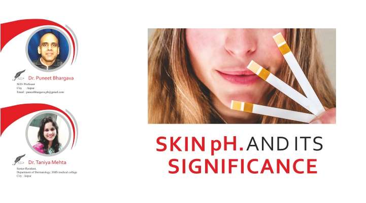
At home, basic pH test strips can be used to measure skin pH. Colour coded scales are available on the strip to assess the pH. Darker the colour, more the pH is in alkaline range and vice versa. Instantly checking pencil kits are also available to check surface pH.

Skin pH levels can be measured by using a pH meter. Guidelines on ideally measuring skin surface pH have been formed in the 5th International conference on Occupational and Environmental Exposure of skin to chemicals. The meter involves a glass H+ sensitive electrode and a reference electrode in a single chamber connected to a voltage meter. It is a very quick and reliable method to measure the pH in a clinical as well as non-clinical setting. Probe is planar and gives single decimal results for easy reading. Interface of distilled water is used for optimum measurements. The commercially available glass pH meters are:

  1. pH meter 1140 (Mettler Toledo)
  2. skin pH meter 900 or 905( Courage and Khazaka)
  3. Russell ph limited
  4. pH meter, Denmark.
pH and its Clinical Significance

Atopic dermatitis: Changes are seen in skin pH in patients of AD. It is believed that patients with AD have decreased secretion of urocanic acid, lactic etc, and impaired lipid synthesis leading to elevated pH, further impairing the barrier function. Loss of function mutations in filaggrin lead to decrease in phospholipase and polycarboxylic acid levels leading to loss of buffering system, hence causing elevated pH in atopic patients. Elevated pH leads to decreased ceramide levels and increases predisposition to Staphylococcal infections which favour a more basic mileu. In atopic patients elevated pH is found all over the body especially lesional skin. Infants show elevated pH on cheeks, a common site for AD lesions in that age group.

Dry Skin and Eczema:

Eczematous and xerotic areas tend to have elevated pH which leads to impaired barrier, added infections and increased skin sensitivity.

Diabetes Mellitus:

DM is a disease in which pH measurements have shown conflicting results. A study showed decrease in pH in type 1 DM and correlating with high glycemic index. Researches have also shown decrease in pH levels in skin of diabetics, especially in intertriginous areas. This may be attributed to autonomic dysfunction causing impaired sweat secretion and low phospholipase levels. Elevated pH maybe the cause of increase in Staphylococcal and candidial infections in diabetics. Candida sp. shifts into hyphal form at basic pH which is pathogenic form, thereby leaqding to candidial intertrigo in diabetics. Skin in these patients tends to be dry, xerotic and also prone to eczematous dermatitis.

Acne

Elevation in skin pH predisposes to acne. Low lactic acid levels and increased pH impairs the barrier function and favors multiplication of Proponibacterium acnes. Studies have been done supporting the above hypothesis. Excessive and repeated washing of face strips away the seboprotective layer too. The use of alpha and beta hydroxy cleansers in acne patients helps maintain an acidic pH.

Soaps, Cleansers and Detergents:

Soaps and cleansers can be categorised as acidic and basic or neutral pH category. Basic and neutral cleansers and soaps can make skin pH alkaline and predispose to its aftereffects as discussed above. Most soaps have a pH within the range of 9-10 and shampoos have a pH between 6-7. The soaps and shampoos used commonly have a pH outside the range of normal skin and hair pH values. Therefore due consideration should be given to the pH factor before prescribing soaps in patients with acne prone or sensitive skin. A single wash by an alkaline soap can shift the pH to the range of 6 to 7 and can take 10-12 hours to revert to normal. Repeated and aggravated washing can further worsen the situation by stripping the sebum from skin surface. Use of mildly acidic, sodium laureth sulphate free cleansers should be advocated in such patients. Similarly in patients with sensitive and irritated skin neutral pH cleansers should be advocated to prevent further aggravation.

Diet and Skin pH

Recent emphasis is on an alkaline diet to improve bone health, muscle growth, Vitamin D levels and overall health of our body. But our skin requires an acidic pH which cannot be maintained by a diet rich in acidic foods. Stressing on neutral and acidic skin applications and washes and maintaining cutaneous hydration can go a long way in maintaining a desired cutaneous pH.

Suggested Readings:

  1. Firooz A, Zartab H, Sadr B, et al. Daytime Changes of Skin Biophysical Characteristics: A Study of Hydration, Transepidermal Water Loss, pH, Sebum, Elasticity, Erythema, and Color Index on Middle Eastern Skin. Indian Journal of Dermatology. 2016;61(6):700. doi:10.4103/0019-5154.193707.
  2. Stefaniak AB, du Plessis J, John SM, et al. International guidelines for the in vivoassessment of skin properties in non-clinical settings: part 1. pH. Skin Research and Technology. 2013;19(2):59-68. doi:10.1111/srt.12016.
  3. Rippke F1, Schreiner V, Doering T, Maibach HI. Stratum corneum pH in atopic dermatitis: impact on skin barrier function and colonization with Staphylococcus Aureus.Am J Clin Dermatol. 2004;5(4):217-23.
  4. Tyebkhan G. A study on the pH of commonly used soaps/cleansers available in the Indian market. Indian J Dermatol Venereol Leprol 2001;67:290-1

*-*-*

Q Switched Ndyag: The Wonder Laser Taking The Aesthetic Dermatologist From Impossible To Possible

The field of laser medicine and surgery has evolved a great deal over the past few years and in my opinion, one of the lasers first discovered and researched extensively upon was the Q switched ND YAG and now we have the nano second Q-switched available in most clinics and many of these are USFDA approved. The one which I work with is the helios 3 and in this article I shall showcase and discuss the various indications both off label and US FDA approved which i have been using this laser for namely:

  • White Hair Reduction
  • Congenital Naevis Removal
  • Atrophic Scar Treatment
  • Scar Texture Improvement
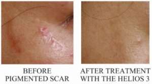
  • Pigmented Scar Revision/Reduction
  • Melasma
  • Atypical Lhr

Now, before I start with the actual details I would like to also emphasize on the US FDA and some finer points which all laser surgeons should follow :-

  • We should always work on demonstrable and reproducible results with definite protocols.
  • The idea is todemonstrate the superiority of lasers over conventional excision measures of scar revision and naevus surgeries.
  • Why US FDA emphasis?? The simple answer is safety and litigation preparedness, It might be surprising to many that whenever in consumer courts the laser in question has been the gold standard US FDA approved, the view of the courts has been most lenient towards the laser surgeons.

q switched ndyag

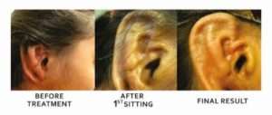
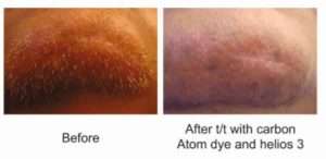
ATYPICAL LHR REMOVED WITH THE QUASI LONG PULSE (NEO GENISIS MODE) HELIOS 3

q switched ndyag

q switched ndyag

POST CHICKEN POX SCARS TRETED WITH THE HELIOS 3

Rationale of Neogenesis, Quasi Long Pulse Mode

  • This mode utilizes the microsecond pulse instead of the nanosecond pulse, hence dermal collagen synthesis is triggered and dermal remodeling takes place.
  • Since the spot size can be varied linear and odd shaped scars can be treated successfully.
  • Texture and tone can also be dealt with Helios 3 so the nanosecond mode with the zoom and fractional handpiece is absolutely safe with a pulse width of 8 it is ideal to treat hyper-pigmented and pigmented scars.
GREY/WHITE HAIR LHR

q switched ndyag 3

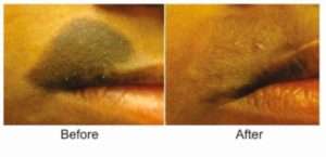
Treatment of Congenital Naevus (Ablative Indications)

GREY/WHITE HAIR LHR

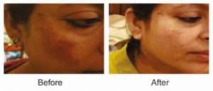
Melasma Treatment Single Sitting Helios 3

q switched ndyag 5

Rationale In Melasma Treatment

  • Once synthesized melanin is packed in melanosomes, melanosomes are then transferred to keratinocytes from dendritic processes ,here is where Helios 3 comes in thus preventing the incorporation to the keratinocyte ,by photoacoustic effect ,as melanosome is heated steam is formed =whitening clinically = endpoint in most cases.
  • The Helios 3 or the fractional Q switched ND YAG works on melanosomes and their suppression and via this we perform the laser dendrectomy, earlier and till today fractional photothermolysis with diode 1550 nm is the US FDA approved protocol however nanosecond and picosecond lasers are showing promising results.
  • Since we all know the nature of melasma is notorious for recurrence despite after all available treatments so the Helios 3 offers a safe and effective way to keep melasma in check in a clinically safe environment
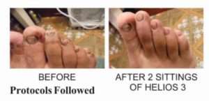
Resistant Onychomycosis /Toe Nail Fungus Treated with the Helios 3

q switched ndyag 6

  • For the Q switched ND YAG,  I use zoom hand piece one to two passes, spot size 3 to 5 depending on size of scar and hyper pigmented area energies 450 to 800 frequency 1 to 3.
  • For linear scars weekly sittings of FR mode zoom handpiece spot size 3, 4 and 5 energies between 1400-1500, energy density up to 7 j/cm2.
  • Duration of treatment in combination will range from 4 to 8 sittings again dependent on the scar characteristics. Usual time gap between 2 sittings is 21 to 28 days. PIH cases during treatment do require fortnightly follow ups.
Conclusions and Summary
  • In scar revision with the Helios 3, we can counter the PIH plus the texture can be improved with Helios 3.
  • In today’s practice, patient expects perfection from his laser surgeon so finer aspects and fine tuning are the norm which can be achieved with the certified prowess of the Helios 3
  • In pigmentory disorders there is near zero down time, freedom to give packages, economical sittings, hope against hope as melasma is considered untreatable conventionally, we can always try and get another chance as Helios 3 is quite versatile.
  • In LHR, we can dare to treat difficult areas inaccessible to the diode tip, ear lobes, grafted areas and white/grey hair can be treated.

*-*-*

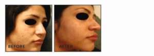
Carbon Peeling

Carbon Laser Peeling or so-called Hollywood Peel was originally discovered in Asia. Because of being the leading anti-aging treatment in Los Angeles, it is called Hollywood Peel. It is gradually growing in popularity in India because of its zero downtime and instant results.

It does not use any harsh, invasive tools, which means no bleeding, adverse reaction or recovery time making it a favourite procedure for Angelina Jolie and Kim Kardashian before red carpet.

Mechanismofaction:

Laser energy lightly absorbs carbon lotion, heating dermis which results in stimulating the building up of collagen leading to tightening and younger looking skin and lightly ablating the top layer of epidermis. Laser energy breaks up pigment that is carried away by blood vessels and lightening effect is achieved. It has bactericidal effect on Propionibacterium acnes and decreases the size of sebaceous glands so a favourable response is seen in acne prone patients.

Indications
  • Enlarged or dilated pores
  • Oily and acne prone skin
  • Presence of fine lines
  • Dull complexion or dyschromia
Contra-indications
  • Acute or chronic inflammatory dermatoses
  • Pregnancy
  • Malignancy
  • History of keloidal tendency
Technique of Laser Carbon Peel

1 Cleansing: It is done with a mild cleanser so as to remove any oil, dirt and makeup and apply eye protective shield.

2 Carbon Lotion Application: Then a uniform layer of medicated carbon lotion is applied over the area to be treated. Then dab it with a cotton pad so as to remove excess of carbon.

3 Laser Pass: We use Q-switched Nd-YAG 1320nm at maximum fluence tolerable to patient as it should not hurt throughout the procedure. The laser pass is given without overlapping. The carbon particles are absorbed by laser and leave skin refreshed and toned.

4 Dressing: The remaining carbon particles, if anywhere, should be cleaned with micelle solution and a layer of vitamin c and hyaluronic acid serum is applied followed by sunscreen.

Post-procedure Instructions
  • Avoid sun and sauna bath for 7 days
  • Use moisturizer and sunscreen on a regular basis.
  • Maintain hydration
Side Effects
  • Erythema and transient swelling which may be present for a few hours and can be managed well with cool packs
  • Post inflammatory hyperpigmentation may also occur
  • Laser burn is quite rare
Results:

Even after a single session, smooth and even skin is noticed. For ideal results, 5-6 sessions at the interval of 2-3 weeks are advised for other indications.

In Summation:

Carbon Laser Peel is a laser procedure that uses a carbon layer as a photo-enhancer to improve the skin’s radiance and promotes a smooth and glowing complexion. This treatment is used to target active acne as the carbon powder penetrates into pores, thus allowing the laser to focus its energy more effectively. It also helps refine enlarged pores to reduce oil secretion, remove open comedones and achieve whitening effects.

*-*-*

Hyaluronic Acid Fillers in Aesthetic Medicine

In our day-to-day life, it is important to look the best that we can, at any age. As we age, our faces lose soft tissue volume and undergo skeletal resorption causing deflation and descent, manifesting in wrinkles and folds. Hence, a lot of people feel what they look on the outside is not what they feel on the inside. Looking young and fresh is becoming important in our social and professional interactions as looking good is associated with positive vibes from others. And it is not sex-specific. Both men and women want to look better and younger. A variety of procedures are available such as botulinum toxin, dermal fillers, chemical peels, lasers and cosmetic surgery. The non-surgical procedures are usually preferred over the surgical ones.

Of the non-surgical treatments, dermal fillers have revolutionized the field of aesthetic medicine. They are increasingly being used by dermatologists and plastic surgeons to diminish or correct the signs of aging. Fillers offer highly gratifying results due to immediate correction of rhytides, lift, enhancement of contours and filling up of hollows.

The first fillers that were approved for clinical use were made of bovine and porcine collagen. However, the results were short-lived, with a potential risk of foreign body reaction with the non-human sources.

By definition the perfect dermal filler should be safe, painless, easy to inject, hypoallergenic, long-lasting and economical. It should also give smooth, natural looking results and feel natural under the skin. Hyaluronic acid (HA) fillers satisfy most of these criteria making them the most common choice of filler and they have now become the gold standard in fillers. Hyaluronic acid binds to water and swells, thereby it volumizes, softens and hydrates the skin. It acts as a matrix for fibroblasts, stimulating the production of collagen which partly contributes to its long lasting effects. Because hyaluronic acid molecule is identical across all species, the risk of allergic reactions is remote. Another advantage of using hyaluronic acid fillers is its ease of correction with hyaluronidase, in case of undesirable outcomes or adverse effects. Most of the newer hyaluronic acid fillers now contain lidocaine, which helps reduce the pain of injection and discomfort of moulding the filler.

HA used in practice today is derived from bacterial cultures of Streptococcus bacteria by biofermentation.

HA fillers can be used to:

  • Plump thin lips
  • Enhance contours eg. of the cheek, jawline and chin
  • Soften facial creases and wrinkles
  • Improve undereye hollows and dark circles
  • Rejuvenating the back of the hands
  • Improving the hydration of skin on the face, neck and hands
  • Hand rejuvenation
  • Non-surgical rhinoplasty

Brands approved for use in India are:

  1. Restylane family of products, which includes Restylane, Perlane, Restylane with lidocaine, Perlane with lidocaine and Restylane vital. This group of products contains particles of cross-linked hyaluronic acid suspended in a non-crosslinked hyaluronic acid gel (biphasic gel). The HA concentration is 20 mg/ml in all the products, but the particle size varies with each product. This makes them one of the stiffest hyaluronic acid fillers. Results last for 6-9 months on an average. Restylane vital is used to enhance the texture and softness of the skin, and is called a skin booster.
  2. Juvederm family of products includes Ultra XC, Ultra plus XC and Voluma XC. Juvederm is a homologous (monophasic) gel with the high degree of cross-linking giving it a smooth consistency. Ultra XC & Ultra plus XC use the Hylacross technology of cross-linking and have 24mg/ml of HA. Their tissue life is about 6-12 months. Voluma XC uses the Vycross cross-linking technology and has 20mg/ml of HA giving it excellent firmness and pliability. The XC range contains lidocaine to reduce the injection pain. The longevity is about 18-24 months.
  3. Esthelis family of products includes Esthelis, Fortelis, Modelis and Mesolis; with HA concentration is 22.5 mg/ml, 25mg/ml, 26 mg/ml &14 mg/ml respectively. Esthelis, Fortelis and Mesolis are approved for use in India. They are monophasic gels with an extra degree of cross linking (cohesive polydensified matrix) except Mesolis which is not cross-linked. Longevity ranges from 6 to 18 months depending on the density of the product.
  4. Emervel lip and Emervel volume have recently been approved for use in India. It has 20 mg/ml of HA, and is biphasic filler similar to Restylane. Results last for 6 to 12 months.
  5. Yvoire classic and Yvoire volume have recently been approved for use in India. The HA concentration is 22mg/ml. It is a biphasic filler with results lasting for 6 to 12 months.

Injection Techniques

They vary from filler to filler. Most common techniques are serial puncture or bolus technique and, linear threading which could be anterograde or retrograde with or without fanning. They can be administered via needle or cannulas. Cannulas are fast gaining popularity among doctors when it comes to treatment of certain areas such as tear trough, cheek, hands. The chances of bruising are less with cannula and larger areas can be treated from a single point, reducing the pain of treatment.

Fillers of varying softness can be placed at different levels in the tissue (sandwich technique) to provide a more natural looking result. Softer fillers are placed more superficially and denser fillers, deeper for a desirable cosmetic outcome.

It is important to be individually trained on the injection techniques of each filler. It is of paramount importance to know the anatomy of the facial skeleton, soft tissues and the neurovascular structures to ensure product placement at the right level and prevent any complications from inadvertent injury.

Considerations in choosing an HA filler:

  1. FDA approval
  2. Cost
  3. Duration of correction
  4. Ease of injection
  5. G prime (G’) or gel hardness – Harder gels like Perlane or Juvedermvoluma are placed deeper in tissue or over the bone whereas softer gels like Restylane, Esthelis or Ultra XC are used on the undereyes and lips.

Adverse effects:

  1. Swelling, erythema & bruising are commonly seen after procedure. These are self-limiting and subside within a week or two. Good injection practices can minimize these events. Use of a cannula has reduced the incidence of bruising.
  2. Pain during the procedure can be minimized by injecting slowly. Products that contain lidocaine reduce the discomfort of the procedure and make molding of the filler quite comfortable.
  3. Tyndall effect: If fillers are injected too superficially they can create a bluish hue due to the Tyndall effect.
  4. Vascular compromise: Although rare, it is one of the most severe side-effects of filler procedure. It can occur due to compression or inadvertent intra-arterial injection of the filler. Compression manifests a few days after the filler procedure with pain at site of injection redness along the site and distribution of the vessels due to opening up of collateral circulation. Intra-arterial injection of filler can result in severe pain and necrosis in the area supplied by the vessel. Treatment includes immediate injection of hyaluronidase in the filler treated area, and vasodilator therapy.
  5. Nodules: Injection close to the surface or large boluses can cause nodule formation. It can also be a delayed phenomenon due to bacterial biofilm formation. They are treated by hyaluronidase into the nodule, with or without antibiotic therapy.
  6. Poor cosmetic outcome can be corrected with hyaluronidase injection.

The demand for soft-tissue augmentation is slowly increasing in India. Newer products with better tissue compatibility and longer lasting results are becoming available. The current aesthetic practitioner will need to be aware of the current availability, application and future potential of dermal fillers.

*-*-*


Melanocytic Naevi: Treatment Modlities

Introduction:-

Melanocytic naevi are common benign skin conditions in Dermatology. Majority of the lesions usually go un-attended unless it causes aesthetic concerns or becomes symptomatic. Proper management of the naevi offers both the patient and the treating physician a high degree of result oriented satisfaction.

Definition:-

Naevi means “birthmark” or “maternal impression”. They are benign clusters of melanocytic naevus cells arising due to proliferation of melanocytes at the epidermis, dermal–epidermal Junction or the dermis.

Etiology:-

Melanocytes are pigment producing cells of the body. Their major function is to produce melanin. Melanocytic naevi are derived form melanoblast cells which are in turn derived from the neural crest cells during embryogenesis. Some of these melanocytes complete their migration towards the basal layer and dermo-epidermal junction while some get arrested in the dermal region. Melanocytes may be found distributed throughout the dermis, around and within the walls of blood vessels, around adnexal structures such as hair follicles, within the subcutaneous tissue and sometimes within skeletal muscle, smooth muscle bundles, nerves, or sebaceous glands. These various locations of melanocytes results in varied types of melanocytic naevi.

Clinical Features:-

Naevi are common cutaneous findings and appear on almost all human skin. They are found to be less frequent at birth and increase in incidence during the first 3 decades of life. Various studies suggest that the number of acquired melanocytic naevi is a factor dependent on genetics and sun exposure. Melanocytic naevi are essentially classified as:-

  1. Congenital melanocytic naevi
  2. Acquired melanocytic naevi

Dermatosurgeons commonly encounter the acquired type of melanocytic naevi for excision or removal usually for aesthetic purposes. The acquired nevi are benign proliferations of melanocytic naevus cells which are biologically stable and develop from birth. However, melanocytic nevi can be found in association with melanoma. The true frequency of transformation of a melanocytic nevus into melanoma is not known and the estimated prevalence varies, with some data suggesting that up to 40% of melanomas are associated with a precursor melanocytic nevus. According to one study the single greatest predictor of risk for developing melanoma is the total number of nevi.

Classification of Acquired Melanocytic Naevi:-

Common acquired melanocytic nevi are generally less than 1 cm in diameter and evenly pigmented. Some atypical melanocytic nevi exceed 1 cm in size, especially when such lesions occur on the trunk. Depending on their location on the skin, they are classified as below:

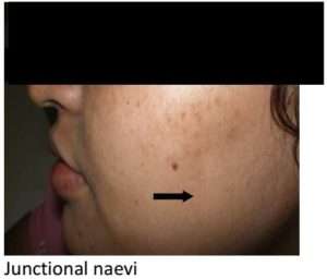
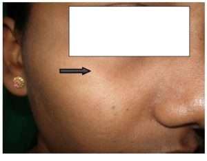
  • Junctional melanocytic nevi are generally macular and typically range from brown to brownish-black.
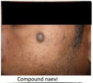
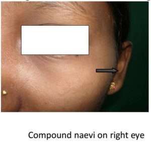
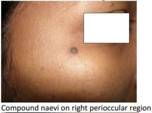
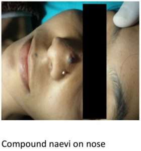
  • Compound naevi are benign proliferations of melanocytes in the epidermis, showing evidence of migration of cells into the dermis and ‘maturation’ of those cells within the deeper dermis. Hence they have both epidermal and dermal components.
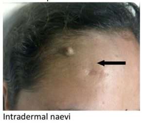
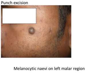
  • Intradermal naeviare benign tumours of melanocytes in which there is no longer epidermal proliferation; the naevus cells have migrated into the dermis and mature there. They are generally skin coloured lesions. Compound and Intradermal melanocytic naevi display elevation relative to surrounding uninvolved skin and hence present as papules or nodules.

Management

Treatment for acquired melanocytic naevi is usually sought for two reasons:

  1. Diagnostic purpose to rule out malignancy
  2. Therapeutic purpose for improving aesthetic appearance

The development of a new area of pigmentation within a long-standing non-pigmented or lightly pigmented compound or Intradermal melanocytic nevus is a cause for concern. While pigmentary changes could be due to incidental inflammation or recent irritation or trauma, the possibility of evolving melanoma cannot be ignored.

Investigations:-

The patient has to be subjected to routine blood tests and histopathological evaluation as follows and send the excised specimen for histopathological examination to rule out malignancy:

  • Complete blood count, platelet count BT, CT
  • Blood sugar
  • Serum Creatinine
  • VDRL
  • Hbs Ag
  • HIV

Pre-Procedure Counseling:

  • Patient has to be properly counseled and explained This helps in better compliance and a better patient doctor rapport and lesser medico-legal issues for the operating doctor.
  • Pre-operative photographs and an informed consent form duly signed by the patient forms an integral part of the pre-op preparation.
  • Physical fitness of the patient should be assessed, if a known case of diabetes or hypertension, necessary precautions should be taken.
  • If on any medications like NSAID or aspirin, it should be discontinued for 2 weeks before the date of procedure.

Procedure:-

Local anesthetic is infiltrated around the naevi. Plain lignocaine or with adrenaline is used accordingly.

Topical EMLA cream can also be used in cases of Junctional naevus. Once the area is anesthetized, cleansing and scrubbing is done and draped.

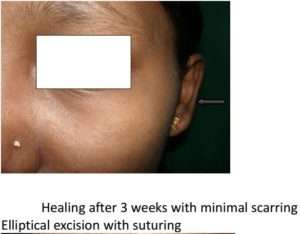
ELLIPTICAL EXCISION:-

Here, the naevi is excised from the skin in an elliptical manner keeping the incision aligned along the RSTL and later this is sutured in bi-layered manner to achieved better cosmesis. This method has various advantages and is preferred when the naevi has to be subjected for biopsy in suspected cases of malignancy.

Procedure:

  1. Instruments required: Surgical bard parker no 15, Adson’s forceps, skin hook, undermining scissors, surgical marker, needle holder, sutures including both intradermal and skin, sizes of which differ according to the area of the skin being operated upon. Usually prolene and vicryl is preferred.
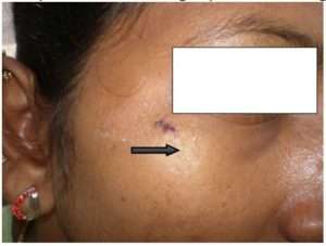
  2. With a surgical marker the naevi is marked, taking care to align it along the resting skin tension line.
  3. With a surgical blade no 15 incision is made along the marked region, cross hatching is avoided.
  4. With a toothed forceps the edge is lifted and skin with the naevi is excised with the surgical blade.
  5. The edges are undermined with an undermining scissors.
  6. Intradermal sutures are put with absorbable sutures
  7. Skin closure is achieved with non absorbable sutures.

Advantages: –

  • Surrounding normal skin can also be subjected for biopsy
  • Depth of the lesion can be controlled
  • Suitable for detecting suspected cases of malignancy
  • Aesthetically better result since lesion excised along RSTL
  • Excision aids in both therapeutic and diagnostic applications

Disadvantages:-

  • Surgical skill needed, since it involves suturing
  • Chances of recurrence if malignant lesion is not excised completely
  • Small to medium sized moles can be removed and sutured, larger area, if excised requires skin graft for closure.

Indications: –

  1. Diagnostic purpose: To rule out malignancy
  2. Therapeutic purpose: Excision of naevi which are suspicious and for aesthetic purposes.

Contraindications: –

  1. Active infection, keloidal tendency, bleeding diathesis, diabetes.
  2. Lesions around important structures like eyes and mouth, joints etc should be avoided since it results   in contractures
PUNCH EXCISION:

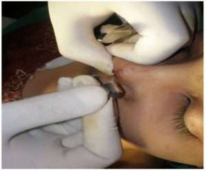
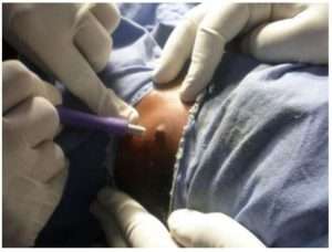
This technique is performed with the help of a metallic cylindrical punch with sharp edges. The punches are selected depending upon the size of the lesion and are available from 1 mm to 5 mm sizes.

Instruments: – disposable or autoclaved punches, skin hooks, undermining scissors, skin sutures.

Procedure:

  1. A disposable punch is selected according to the size of the naevi.
  2. Skin is stretched between the index finger and thumb.
  3. Punch is driven around the lesion and turned in clockwise direction.
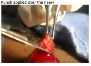
  4. Rotation of the punch is stopped when there is a feel of giving away.
  5. The naevi, included in the punched out area will appear to float in the surrounding skin.
  6. Punched out lesion is lifted with a needle and the base is cut from the surrounding skin with scissors.
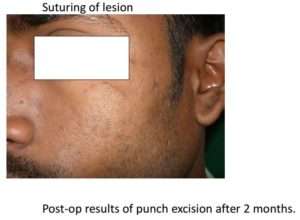
  7. Hemostasis is achieved if the size of naevi is small and skin is left to heal by secondary intention.
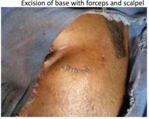
  8. If the lesion is larger, skin closure is achieved by using non-absorbable sutures.

Advantages:-                 

  • Easy to use since it requires less skills.
  • Requires lesser time.

Disadvantages:-       

  • May result in dog-ear during repair of the circular defect on skin.
  • Blind procedure, hence depth of the lesion excised cannot be controlled, resulting in recurrences.
  • In suspected cases of malignancy, perilesional skin is not available for histopathological evaluation.

Indications:-      

  1. Elevated naevi
  2. Useful in lesions involving subcutaneous tissue.

Contraindications:-

  1. Active infection, keloidal tendency, bleeding diathesis, diabetes.
  2. Suspected cases of malignancy.
  3. Friable and fragile tissue
SHAVE EXCISION

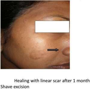
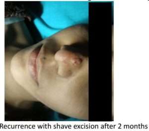
A technique where in the elevated lesion is shaved from the skin using either a surgical blade or sterilized razor blade. The depth of the lesion removed can be controlled leaving the dermis intact hence resulting in minimal scarring.

Instruments: – Sterilized razor blade or no 15 bard-parker surgical blade.

Procedure:

  1. The lesion to be excised is held and stabilized with the index and thumb finger of the left hand.
  2. The blade is held with the right hand parallel to the surface of the lesion.
  3. With a to and fro movement maintained along the base of the lesion, it is sawed in-toto and separated from the base.
  4. The lesion is released from its base and remains on the blade, thus any remaining attachment is cut with the help of scissors.

Advantages:-

  • Easy to perform, lesser skills required.
  • Scarring is lesser since the dermis is left intact resulting in good aesthetic results.

Disadvantages:-

  • Cannot perform in cases of suspected malignancy.
  • Difficult to perform if lesion is not intact.
  • If the depth is more may result in scarring.

Indications:-

  1. Lesions on concave areas of body surface.
  2. Elevated naevi.
  3. Excisions where aesthetic outcome is a priority.

Contraindications:-

  1. Active infection, keloidal tendency, bleeding diathesis, diabetes.
  2. When malignancy is suspected.
  3. Flat lesions.
RADIOSURGERY:-

Commonly performed office procedure for excision of lesions, based on thermal ablation and destruction of tissue with a frequency of alternating current in the range of radiowaves. To perform the procedure a radio surgical unit with tungsten electrodes is required. With this unit, cutting of the target tissue, excision along with control of blood loss through homeostasis is achieved.

Procedure:-

  • The electrode is selected according to the target tissue and power intensity is adjusted .Target tissue is marked and the electrode angled perpendicular to the lesion and incision is made along the margins of the lesion.
  • With an Adson’s forceps the tissue is grasped and the electrode is forwarded beneath the tissue in case of a small lesion. Flat lesions are approached form above downwards.
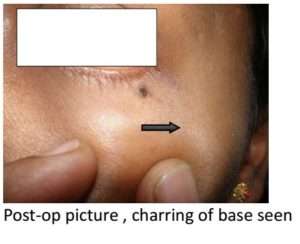
  • The tissue is excised and the remnants are carefully removed, taking care to differentiate between naevus tissue and charred normal skin.
  • Homeostasis is achieved by pressure or using the option of coagulation.
  • Depth of the lesion removed should be controlled since it may result in scarring.

Advantages:-

  • Easy to perform , office procedure
  • Less time consuming,
  • Lesser tissue damage, thus resulting in fewer complications.
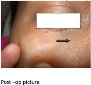
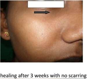
  • Better option for excision of lesions on thinner skin like eyelids.

Disadvantages

  • Larger lesions may result in scarring
  • Cannot be used in cardiac patients.
  • Recurrences occur if not excised adequately.
  • Pigmentary disturbances post procedure may occur.

Indications:-

  1. Flat lesions and small elevated lesions.
  2. Lesions around the structures of the face like – eyes, mouth and nose.

Contraindications:-

Active infection, keloidal tendency, bleeding diathesis, diabetes

  1. Patients suffering from cardiac disease, with pacemakers, metallic prosthesis.
  2. Use of spirit or alcohol based cleansers.
LASER SURGERY:-

Ablative lasers such as Co2 and Erbium: Yag have been used successfully for surgical removal of moles. Though not used very commonly, laser surgery is preferred since it can be used in cardiac patients and is less painful, provides an almost bloodless field since there is coagulation of blood vessels. Local anesthesia is avoided hence all the side –effects and complications associated with local anesthetic.

The cost of these procedures along with the chances of post-inflammatory pigment disturbances are the disadvantages with this procedure.

Conclusion:-

Naevi are common skin conditions which usually do not warrant any treatment. However, in some tradition, presence of naevi in certain sites is regarded as inauspicious. Many patients seek treatment to overcome this superstition. Seeking treatment for cosmetic purposes is also on the rise. At present, there is choice of various modalities of treatment available, and it is the duty of a dermatosurgeon to provide the best possible option to a patient. With proper selection the right procedure, good cosmetic results can be achieved.

The authors have tried various modalities of treatment for mole removal and opine that excision and suturing along RSTL is the best option available as it offers good cosmetic results. However the choice of a procedure may be discussed with the patient and decided as per the patients needs.

A recent study conducted by the King’s college of London states that in a case, a series of patients with more than 100 moles had lesser instances of osteoporosis and fewer wrinkles. So the day may not be far away when cases of mole removal for cosmetic reasons may actually decline and moles may be perceived as good omen!

naevi

naevi

naevi

naevi

naevi

naevi

naevi

naevi

naevi

naevi

naevi

naevi

naevi

naevi

naevi

naevi

naevi

naevi

naevi

naevi

naevi

naevi

naevi

*-*-*

Racoon Eyes- A Nightmare!

Periorbital Hyperpigmentation (POH)

is one of the most commonly encountered conditions in routine dermatology practice. Pigmentataion around eyes is an identity for animals not for humans! Dark circles under the eyes (DC) are defined as bilateral, round, homogeneous pigment macules on the infraorbital regions. DC are caused by multiple etiologic factors that include dermal melanin deposition, postinflammatory hyperpigmentation secondary to atopic or allergic contact dermatitis, periorbital edema, hormonal variations, drugs, superficial location of vasculature, shadowing due to skin laxity, wrinkles and tear trough. They can be genetic and can be seen as an extension of facial pigmentary demarcation lines and rarely due to facial acanthosis nigricans.

Management

  1. Lifestyle modification
  2. Dietary modification
  3. Topical Medicaments
  4. Chemical Peels
  5. Lasers
  6. Others
Lifestyle Modification:
  • Adequate water intake.
  • Avoidance of stress, regular exercise, meditation
  • Adequate supply of quality sleep
  • Avoidance of smoking: constriction of blood vessels due to smoking prevents adequate blood flow to the skin under the eyes thus triggering dark circles.
  • Application of sunscreens over periorbital area.
  • Wear Hats with wide brims.
  • Wear dark glasses.
  • Use make-up containing SPF
  • Avoid make-up that feels too heavy and sticky with fragrance
  • Keep a check on the triggering factors(if any) – eye drops, facial creams, hair dye etc
  • Abstain from rubbing your eyes- repeated rubbing and scratching leads to breakage of delicate capillaries in the area and results in dark circles
  • Avoid giving too much pressure while removing make-up to avoid exacerbation due to rupture of those capillaries
  • Keep check on the expiry dates of your eye cosmetics
  • Follow proper application method of creams: gentle strokes from outside inwards on lower lids and inside outwards on upper lids.
Dietary Modification:
  • Consume diet rich in beta-carotene, lycopenes, Iron, antioxidants, minerals like selenium and Vitamin A, C and E.
Topical Medications:
  • Sunscreens
  • Creams containing very mild Hydroquinone, Alpha hydroxyl acids, Vitamin c, Retinols (low concentrations), Vitamin K, Vitamin E, Collaging boosters and Antioxidants
  • Skin lighteners like kojic acid, Azelaic acid, Mandelic acid, Arbutin, Tetra hydro curcumin,Topical Vitamin C
  • Short term use of triple combination creams-Kligmans formula

Note: Creams containing Retinols, Vitamin C and Hydroquinone are particularly unstable products and if they turn brown in colour or if Vitamin C products crystallize, it is advisable to discard them.

Chemical Peeling:

Arginine peel

Lactic acid peel

Glycolic acid

TCA 3.75%

Citric acid

Retinol Peels

Lasers:

Q Switched Nd YAG laser

CO2 LASER

Q-Switched Alexandrite 755 nm

Ablative laser resurfacing.

Others:

Hyaluronic acid fillers – tear trough

Botulinum Toxin – crows feet correction for a refreshed look

PRP

Mesotherapy

Autologous fat transplantation

Blepharoplasty

Carboxytherapy

*-*-*