Laser Treatment of Acne Scars

Introduction

Acne scarring presents a challenging aesthetic problem to the aesthetic practitioner. Acne is defined as a chronic inflammatory condition of the pilosebaceous glands. It is experienced by 80% of individuals between the age group of 11-30 years. However the severity, age of onset and duration vary widely. [1]

Acne lesions can lead to scarring due to imbalance of matrix degradation and collagen biosynthesis during the process of wound healing. [3]

Aggressive treatment of acne at an early stage prevents scarring. But once acne scarring has occurred, the physician and the patient are left to struggle with the different available options for improving the appearance of the skin. [1]

Acne scars are categorized as:

  1. Hypertrophic
  2. Atrophic

Atrophic acne scars can be further classified as:

  1. Ice pick
  2. Rolling
  3. Boxcar

The boxcar type can be further divided into deep or shallow. As such there is some variation with all the types. This classification system is developed keeping in mind the practicality and therapeutic options available for treatment. [1, 3]

Hypertrophic acne scars are described as raised, erythematous and firm nodular lesions whose growth is limited to original tissue injury site.

Ice pick scars are deep, narrow (<2mm) and sharply marginated epithelial tracts extending vertically to the reticular dermis or up to the subcutaneous tissue. [1]

Rolling scars are wider usually 4-5 mm. They occur as a result of dermal tethering of otherwise normal appearing skin. [1]

Boxcar scars are sharply delineated epithelial tracts that extend into the dermis but, unlike ice pick scars do not taper at the base. [3]

Histopathologically, acne scarring destroys the underlying collagen which makes it difficult to treat. Many treatments have been tried and tested in past with varying degrees of success. Some of which are dermabrasion, chemical peels, Subcision, punch excision followed by suturing or grafting, micro-needling and dermal fillers. [2, 3]

Nowadays with advancement in the laser technology and availability of new resurfacing lasers, it is becoming easy to treat this therapeutically difficult condition. [3] Lasers allow for direct smoothening, collagen neogenesis in the scarred skin and contraction of acne scars. These lasers far exceed the efficacy of traditional treatment.

History of Laser Resurfacing

Lasers were there in the market since long but the field was revolutionized by Anderson and Parrish in 1983 [4] when they elucidated the principles of selective photothermolysis. In order to achieve maximal target destruction and minimal damage to the surrounding tissues the wavelength of laser, pulse duration and fluence each must be carefully chosen. [3]

The Pulsed Dye Laser (PDL) was the first reported laser for scar improvement in the year 1993. [5] Following this and with the advancement in the laser technology the laser scar revision has tremendously progressed. Several qualities of scar influence the choice of laser wavelength and treatment parameters including the size, colour and texture of scar. [3]

Lasers in Hypertrophic Acne Scars

PDL was the first Non Ablative Laser system being successful in the treatment of hypertrophic, erythematous facial acne scars.

PDL acts by:

  • Reducing transforming growth factor –β expression
  • Deposition of type 3 collagen
  • Proliferation of fibroblasts
  • Thermal Coagulation of collagen fibres
  • Breaking of disulphide bonds followed by Neocollagenesis. [3]

In a study, 10 subjects having shallow to moderately deep facial acne scars, who underwent a single treatment session of 585nm PDL. All 10 subjects showed marked clinical improvement and no adverse effects were reported. [6]

Post treatment purpura which persists for a few days is the commonest side effect following treatment with PDL. Edema which occurs at the treatment site usually subsides within 48 hours. In order to protect the skin, for the first few post-operative days a topical healing ointment can be applied. There should be strict sun avoidance and sunscreens should be used. Patients should be advised to clean the treated areas gently with water and soap on daily basis. [3]

Hence 585nm PDL is the laser of choice in the treatment of hypertrophic erythematous acne scars.

Lasers in Atrophic Acne Scars

Atrophic acne scarring treatment with lasers is a precise and well-tolerated procedure. It shows clinically demonstrable efficacy with minimum side effects. It can also be used in conjunction with other scar treatments.

Ablative Resurfacing:

Two wavelengths for ablative laser resurfacing are:

  1. Infrared CO2 (10,600 nm)
  2. Er:YAG (2940nm)

Target chromophore for both these lasers is water. Hence intracellular tissue water absorbs the emitted energy which leads to heating and vaporization of the superficial skin. [3]

These lasers work by causing shrinkage of collagen, neocollagenesis and collagen remodelling which leads to marked improvement in acne scarring. [3]

The prolonged histological and clinical effects of high energy pulsed CO2 laser was documented in 1999. In this prospective study which included 60 patients, continued clinical as well as histological improvement showing dermal remodelling and progressive neocollagenesis was noted for 18 months after resurfacing with CO2 laser. [7]

When the Er: YAG (2940nm) laser is compared with the CO2 (10,600nm) laser, one can observe that 2940nm wavelength is more efficiently absorbed by water. Er: YAG has much shorter pulse duration than the CO2 laser. But since Er: YAG causes limited thermal injury to the skin compared to that of CO2 the amount of collagen contraction is also less. [3]

To overcome these limitations, modulated Er: YAG and combination Er: YAG-CO2 lasers were developed. These lasers emit combination of short ablative pulses as well as long coagulative pulses. The result was improved haemostasis and increased collagen shrinkage and remodelling. The modulated Er: YAG was thus similar in efficacy to that of CO2 lasers. [3]

In a study that assessed the outcome of different Er: YAG lasers in different types of acne scarring in 158 patients, showed improvement in ice pick and boxcar (shallow) scars. Long pulse Er: YAG laser showed improvement in rolling and boxcar (deep) scars. [8]

Though Ablative Laser Resurfacing for acne scars is an OPD procedure careful history taking, consent, clinical photographs, preoperative counseling particularly regarding the post procedure recovery period is required. [3]

Absolute contraindications:

  • Active viral, fungal or bacterial infection
  • Inflammatory skin condition (e.g. Psoriasis, eczema, etc.)
  • History of keloids
  • Use of oral retinoids within the preceding 6-month period

Potential complications post laser scar resurfacing includes dermatitis or infection, which usually occurs during the 7-10 days re-epithelialization process. Prophylactic antibiotics/antibacterial reduces the risk of infection. [9-11]

When aggressive laser parameters are used, it may lead to complications such as ectropion and hypertrophic scarring. [3]

Non Ablative Resurfacing

Most widely used non-ablative systems are:

  1. 1320 nm Nd: YAG
  2. 1450 nm Diode
  3. 1064 nm Nd: YAG

These lasers deliver deeply penetrating infrared wavelengths with simultaneous epidermal cooling. This leads to neocollagenesis through dermal heating while maintaining the integrity of the epidermis. [12]

The 1320nm Nd: YAG laser has shown significant improvement in atrophic acne scars reported by both the patients and physician without evidence of notable adverse effects. [3]

The 1450nm diode laser showed modest improvement in atrophic acne scarring after 4-6 sessions. [3] In a spilt face study that compared the effects of 1450nm

diode with 1320nm Nd: YAG, after 3 monthly sessions, both lasers demonstrated a mild improvement, but there was greater clinical effect with the 1450nm diode laser. There were no adverse textural changes with both the lasers. [3]

Atrophic acne scarring treated with 1064nm Nd: YAG showed mild to moderate improvement, which was confirmed histologically. [3]

Though the results achieved by non-ablative laser systems are inferior in comparison to ablative one, the low adverse profile of these lasers compensate for their reduced clinical efficacy.

Fractional Laser Resurfacing

Fractional Lasers can be of two types:

  1. Non Ablative Fractional Lasers (NAFL)
  2. Ablative Fractional Lasers (AFL)

Fractional lasers work by creating Microscopic Thermal Zones (MTZ). MTZ are microscopic non-contiguous columns of thermal energy in the dermis. The tissue surrounding each MTZ remains intact which results to quick healing. [3]

There is also improvement in superficial dyspigmentation because NAFL causes gradual exfoliation of the epidermis. This is an added advantage of NAFL over ablative lasers. NAFL requires a series of treatment to achieve optimum clinical improvement. [3]

Many studies have shown clinical improvement of at least 50% or more in atrophic acne scarring after a series of three consecutive sessions. [3]

The consensus guidelines for NAFL for atrophic acne scarring for different types of skin are: [3]

Fitzpatrick skin types I–III

  • Energy – 30-70mJ
  • Level of treatment –  7–11
  • Number of passes – 8-12

Fitzpatrick skin types IV-VI

  • Energy – 30–70 mJ
  • Level of treatment – Low treatment density to avoid Post-Inflammatory Hyperpigmentation
  • Number of passes – are also a few

Ablative Fractional Lasers, in addition to creating MTZ, vaporize the Stratum Corneum. [3] So the post procedure appearance is similar to that of an ablative laser. There have been numerous studies regarding the success of AFL in treating atrophic acne scars. [3]

The ideal patients for fractional laser resurfacing are patients with Fitzpatrick skin types I, II, and III but Fitzpatrick skin types IV–VI patients can also be treated. [3]

While treating patients with AFL, adequate patient education and preoperative assessment are necessary in order to avoid pitfalls and maximize patient outcomes. With fractional laser technology, the chances of pigmentary alteration and unexpected scarring are much less when compared with that of fully ablative lasers. [3]

The energy settings will differ depending on what type of laser is used and also on the severity and type of acne scars. Improved clinical efficacy is noted with higher energy settings, but there are also chances of increased adverse events such as: [3]

  • Pain
  • Erythema
  • Postoperative dyspigmentation

Patients undergoing NAFL are advised to use a mild cleanser as well as a moisturizer several times in a day following each treatment session for the first few days. Sun exposure should be avoided during this period. To prevent infection and other complications in patients undergoing AFL, open or closed wound dressing is advised for the first few post treatment days. [3]

Complications associated with fractional laser resurfacing: [3]

  1. Erythema
  2. Periocular edema
  3. Xerosis
  4. Slight darkening of the skin
  5. Acneiform and herpetic eruptions
  6. Post inflammatory hyperpigmentation

Conclusion:

Acne scarring is a common problem and patients mostly resort to treatment for cosmetic improvement. Most of the treatments that were available in the market were inadequate or insufficient in treating acne scarring. In order to overcome this, Laser therapy for scar revision was investigated.

Different types of acne scars are treated effectively nowadays with different types of lasers. 585 nm Pulse Dye Laser is quite effective for hypertrophic acne scarring. Depending on the individual patient circumstances atrophic acne scarring can be treated successfully with Ablative lasers and Fractional Ablative and Non-Ablative laser systems. These lasers work by contouring the scar tissue via collagen contraction and neocollagenesis. The best outcome for laser scar revision is when the characteristics of scars are evaluated thoroughly the treatment is individualized for each patient. And most importantly both the physician and patient have realistic expectations and treatment goals.

References:

  1. Jacob C, Dover J and Kaminer M. “Acne scarring: A classification system and review of treatment options”. Journal of American Academy of Dermatology (2001); 45: 109-117.
  2. Jordan R, Cummins C and Burls A. “Laser resurfacing of the skin for the improvement of facial acne scarring: a systemic review of the evidence.” British Journal of Dermatology (2000); 142: 413-423.
  3. Sobanko J and Alster T. “Management of Acne Scarring, Part I.”American Journal of Clinical Dermatology (2012); 13: 319-327.
  4. Anderson RR and Parrish JA. “Selective photothermolysis: precise microsurgery by selective absorption of pulsed radiation.”Science(1983); 22: 524-7
  5. Alster TS, Kurban AK and Grove GL, et al. “Alteration of argon laser induced scars by the pulsed dye laser.” Lasers in Surgery and Medicine(1993); 13: 368-73
  6. Patel N and Clement M. “Selective Nonablative Treatment of Acne Scarring With 585 nm Flashlamp Pulsed Dye Laser.”American Society for Dermatologic Surgery (2002); 28: 942-945.
  7. Walia S and Alster T. “Prolonged Clinical and Histologic Effects from CO2 Laser Resurfacing of Atrophic Acne Scars.”American Society for Dermatologic Surgery (1999); 25: 926-930.
  8. Woo SH, Park JH and Kye YC. “Resurfacing of different types of facial acne scar with short-pulsed, variable-pulsed, and dual-mode Er:YAG laser.”Dermatologic Surgery(2004); 30: 488-93
  9. Walia S and Alster TS. “Cutaneous CO2 laser resurfacing infection rate with and without prophylactic antibiotics.”Dermatologic Surgery(1999); 25: 857-61
  10. Alster TS and Nanni CA. “Famciclovir prophylaxis of herpes simplex virus reactivation after cutaneous laser resurfacing.” Dermatologic Surgery(1997); 25: 242-6
  11. Beeson WH and Rachel JD. “Valacyclovir prophylaxis for herpes simplex virus infection or infection recurrence following laser skin resurfacing”. Dermatologic Surgery(2002); 28: 331-6
  12. Friedman PM, Skover GR, Payonik G, et al. “3D in-vivo optical skin imaging for topographical quantitative assessment of non-ablative laser technology”. Dermatologic Surgery(2002); 28: 199-204.

Increasing Rate of Various Pigmentary Disorders

A Retrospective Study Analysis of Data Trend of Erythema DyschromicumPerstans (EDP) Versus Lichen Planus Pigmentosus (LPP) Versus Post Inflammatory Hyperpigmentation (PIH) seen At DISHARC (DI Skin Health & Referral Centre) Nepal.

Introduction

In the recent years, there has been an increase in pigmentary disorders. Among those, hyperpigmentation is also quite common. When a patient visits dermatologist’s clinic with hyperpigmentation there are certain ways of asserting for the diagnosis. There are few hyperpigmentary disorders which are similar clinically and their proper diagnosis is required and further diagnostic skin punch biopsy is done as the treatment modalities, disease progression and several other factors vary. Nowadays, at the OPD clinic suspected cases of EDPvs. LPP vs. PIH are increasing. Hence a retrospective study of data was done to evaluate the various aspects of these pigmentary problems at DISHARC.

Discussion

Erythema dyschromicumperstans (EDP) is also known as ashy dermatosis. It is a benign, uncommon skin condition that presents with generalized ashy-grey colored macules on the body1. It affects both males and females, and is seen most commonly before the age of 40. Lesions still may be seen in young children and in the elderly.

Most patients are asymptomatic although some may have itching. On clinical presentation, the skin lesions are round, oval or sometimes irregularly shaped macules and patches that are grey, or bluish-brown in colour. They develop in a symmetrical pattern and can reach up to 0.5-3 cm in size. These lesions usually begin on the trunk and spread to the neck, arms and sometimes the face. Early lesions may have an elevated, non-scaly, reddish border and older lesions may have a lighter border. Mucous membranes are not affected.

A biopsy may be performed to rule out other diseases and skin conditions. There are a few therapeutic options for EDP but none that have been shown to be consistently effective. Clofazimine, an antibiotic with antibacterial and anti-inflammatory properties, and dapsone have been shown to be effective in some patients. Hydroquinone and topical steroids have varying results. Other therapeutic modalities include UV light therapy, chemical peels, griseofulvin, antihistamines, and isoniazid. EDP may persist for years although spontaneous remission may occur, especially in children.

Lichen planuspigmentosus (LPP) is a rare form of lichen planus. Areas that are exposed to sun such as the forehead, temples and neck are most commonly affected. However, the macules and patches may also develop on the trunk or in places where two areas of skin touch or rub together (i.e. the armpit, groin, etc).


dermatalk

Result:

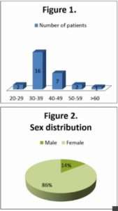
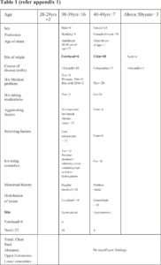
A single centre retrospective study over a period of 6 months was done at DISHARC. Data over a period of 6 months from year 2015 were collected and analysed.

After clinical diagnosis, the suspected cases were advised for skin punch biopsy and histopathological analysis for confirmation of the diagnosis.

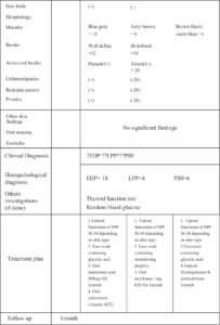
Morphologically lesion colour varied from ashy brown to blue gay and few were violet blue over the common site of origin which was usually over the chin followed by forehead and neck. 12 cases had well defined and 16 cases ill-defined border. There was absence of active red border, lichenoid papules, reticular pattern and none had pruritus and other skin findings. None of the cases were associated with nail, oral mucosa or genitalia findings.

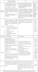
Data were recorded in recording sheets and then evaluated (refer to appendix 1)

EDP-LPP 3

Dermatalk

Dermatalk

Performa EDP/LPP/PIH

Name Age/Sex

Address Profession

History:

Age of onset- Site of origin

Course of disease (months)

H/o any other medical problems –DM/ Htn./Hypothyroidism/ Hyperthyroidism/ Addison’s disease/ Others

H/o taking any medications

Aggravating factors

Relieving factors

H/o using cosmetics

Menstrual history

Clinical examination:

Distribution of lesion-

Localized (affecting one body area)

Generalised (affecting more than one)

Site: Forehead/ Cheeks (Rt./Lt./B/L) Chin/ Nose Symmetrical/ Asymmetrical

Neck Symmetrical/ Asymmetrical

Trunk

Chest Symmetrical/ Asymmetrical

Back (upper/ lower/ both) Symmetrical/ Asymmetrical

Abdomen Symmetrical/ Asymmetrical

Upper Extremities (upper arm/ forearm)

Lower extremities (upper leg/ lower leg)

Skin folds- Submammary folds, axillary, inguinal

Morphology of lesions:

Macules: Blue gray/ ashy brown/ violet blue

Border: well defined/ ill defined

Active red border: Present/ absent

Lichenoid papules:  Present/ absent

Reticulate patter: Present/ absent

Pruritus: Present/ absent

Other skin findings-

Nail findings-

Oral mucosa-

Genitalia-

Clinical Diagnosis

Investigations:

Biopsy:

Hyperkeratosis/ hypergranulosis

Thinned epidermis

Basal cell vacuolization

Perivascular infiltrate

Incontinence of pigment & melanophages

Colloid bodies

Lichenoid infiltrate

Other changes

Histopathological diagnosis:

Other investigations (if done)

Treatment given

Follow up

References:

  1. org/page/Erythema Dyschromicumperstans
  2. 2011 Jan-Feb;9(1):63-4.A case of Cinderella: erythema dyschromicumperstans (ashy dermatosis or dermatosiscinecienta).Muñoz C1,Chang AL.
  3. JIMSA January-March 2014 Vol. 27 No.A Study of 10 Cases of Ashy Dermatosis and Lichen Planus Pigmentosum NeerjaPuri Consultant Dermatologist, Punjab Health Systems Corporation, Ludhiana, Punjab, India
  4. Fitzpatrik dermatology in general medicine Vol8Lowell A. Goldsmith, Stephen I. Katz, Barbara A. Gilchrest, Amy S. Paller, David J. Leffell, Klaus Wolff
  5. Lichen planus.DermNet NZ. March 2015; http://dermnetnz.org/scaly/lichen-planus.html.
  6. J ClinAesthetDermatol. 2010 Jul; 3(7): 20–31.PMCID:PMC2921758 Postinflammatory Hyperpigmentation A Review of the Epidemiology, Clinical Features, and Treatment Options in Skin of ColorErica C. Davis, MDa and Valerie D. Callender, MDa,b

*-*-*

A Smouldering Volcano

Abstract:

Keratoacanthoma is a rapidly evolving tumour of the skin, composed of keratinizing squamous cells originating in the pilosebaceous follicle.[1] It can progress very rarely to squamous cell carcinoma. It affects the sun-exposed area in the sixth or seventh decades. In temperate regions, 70% keratoacanthomas are localized on the face while in the subtropics they involve the arms, dorsa of hands and lower extremities. Surgical excision is a modality of treatment. Overall recurrence rate is 2.4%. [2]

Introduction:

Keratoacanthoma was first described by Sir Jonathan Hutchinson in the 1880s.[3] It affects sun-exposed areas in Fitzpatrick’s skin types 2 and 3. It presents usually as a solitary dome shaped erythematous skin coloured nodule with a central keratin plug. After a rapid initial growth phase, it spontaneously involutes in 4-6 months. Recurrences occur especially when the tumour is present on trauma-prone regions like hands, fingers, lips or ears. There are various variants of keratoacanthoma; out of which giant keratoacanthoma are locally aggressive without metastasis. They are difficult to differentiate clinically and histopathologically from highly differentiated squamous cell carcinoma. In this report, we present a case of giant keratoacanthoma which was treated with surgical excision.

Case Report:

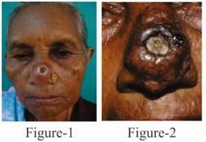
A 56 year-old female patient, farmer by occupation, presented with an asymptomatic rapidly growing tumour on the nose for a year. The swelling started as a nodule which rapidly grew in size with peripheral extension. It further developed raised rolled out margins and a central depression.(Fig 1) The size of the lesion was about 3 cm x 4 cm x 1 cm, located on the bridge of the nose.(Fig 2) It was firm to hard in consistency. There was no bleeding or tenderness on palpation. Differential diagnosis of Cutaneous Leishmaniasis, giant Keratoacanthoma, keratoacanthoma Centrifugum Marginatum and Squamous Cell Carcinoma were considered.

Smouldering

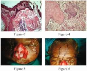
The lesion was surgically excised with a wide margin and wound was closed with flap surgery. (Fig 3, 4, 5)Tissue was sent for histopathology. On histopathological examination after H and E staining, a well circumscribed circular lesion was seen, with a central cup shaped crater which was filled with keratin. (Fig 6) At either side of the lesion, epidermal buttressing was present. A few horn pearls were also seen. (Fig 7) No cells containing LD bodies were seen. Thus, the diagnosis of Keratoacanthoma was confirmed. The patient has regularly consulted us for the last three years, with no evidence of recurrence.

Smouldering 2

Discussion:

A Keratoacanthoma was described by Freudenthal of Wroclaw in the 1940s.[3] Keratoacanthoma develops with an aggressive and infiltrative nature. Keratoacanthomas larger than 20-30 mm are termed as Giant Keratoacanthomas.[4] Less than 50 cases are reported in literature.  It is an acquired disorder with no genetic predisposition. Several factors are involved in the pathogenesis of Keratoacanthoma that includes ultraviolet radiation, smoking, contact with chemical carcinogens like pitch, mineral oil, tar, trauma and vaccination.[5]  Giant Keratoacanthoma  occurs on the eyelid and nose. Our patient had a lesion on the nose. Being a farmer by occupation, exposure to ultraviolet radiation and trauma could be the probable etiological factors.

Progressive peripheral extension with a raised rolled-out margin and atrophy at the centre is a characteristic feature of Giant Keratoacanthoma which was seen in our case. This sort of typical appearance may arise as a result of sequential involvement of multiple hair follicles in a centrifugal fashion.[6]

Giant Keratoacanthoma causes destruction of underlying tissue and spontaneous involution takes place after several months, often accompanied by detachment of a large keratotic plaque.   Keratoacanthoma lesions are benign and regress spontaneously. Locally aggressive nature of Giant Keratoacanthoma creates diagnostic difficulties for the dermatosurgeon as to differentiate from a malignant tumor like Squamous Cell Carcinoma.

Giant Keratoacanthoma has been treated successfully with oral retinoids.[4,7] which should be given until complete clearance of lesion. If surgery is not possible, radiation therapy can be considered. Other treatment modalities successfully used include topical 5- Fluorouracil[9,10], intra-lesional injections of interferon Alpha-2a[4,11], Methotrexate[13], or Bleomycin, or Mohs micrographic surgery.[12]

Surgical intervention should be a preferred mode of therapy as it totally removes the tumor. Being a lesion in an elderly patient on sun-exposed areas and as it was locally aggressive, we opted for wide surgical excision. This achieved the best cosmetic results with minimal scarring.

Conclusion:

Keratoacanthoma is a fast growing aggressive tumor with spontaneous regression. Development of giant variant of Keratoacanthoma with infiltrative and aggressive behavior is known which enables dermatologist to differentiate it from highly differentiated Squamous Cell Carcinoma. Surgical excision like flap surgery is a treatment of choice for Giant Keratoacanthoma.

We are presenting this case for its relative rarity in the Indian scenario and for the good cosmetic results obtained as well as the lack of local recurrence after a number of years of follow-up.

References:

  1. MacKie RM, Quinn AG. Non-Melanoma skin cancer and other epidermal tumors. In: Burns T, Breathanach S, Cox N, editors. Rook’s textbook of dermatology: 8th Singapore: Fabulous Printers; 2010. p.52.42-44.
  2. Larson PO. Keratoacanthomas treated with Mohs’ micrographic surgery (chemosurgery). A review of forty-three cases. J Am Acad Dermatol. 1987; 16:1040–1044.
  3. Sujay khandpur, M.Raman (Authors), IADVL Textbook Of Dermatology,Skin Tumors(chapter 46).Third Edition. (editors) R.G.Valia & Ameet R. Valia. Mumbai Bhalani Publishing House.(1475-1538).
  4. Schwartz RA. Keratoacanthoma: a clinico-pathologic enigma. Dermatol Surg. 2004; 30(2 Pt 2):326–333.
  5. Piyush B Borkhatariya, Shweta Gupta, Damodar Bang, Ranjan C Rawal. Case report and review of literature:Correspondence: Keratoacanthoma centrifugum marginatum. Indian Journal of Dermatology. 2007; 52 (4): 204-205.
  6. Kirkham N. Tumors and cysts of the epidermis. In: Elder DE, editors. Lever’s histopathology of the skin. 10th USA: Lippincott Williams and Wilkins; 2009. p. 791-850.
  7. Schwartz RA. Keratoacanthoma. J Am Acad Dermatol. 1994; 30(1):1–19.
  8. Meghana M Phiske, Ganesh Avhad, HR Jerajani. Case Report: Keratoacanthoma centrifugum marginatum at an unusual site. Indian Journal of Dermatology. 2013; 58 (1): 74-76.
  9. Gray RJ, Medland NB. Topical 5-fluorouracil as primary therapy for keratoacanthoma. Ann Plast Surg. 2000;44(1):82–85.
  10. Parker CM, Hanke CW. Large keratoacanthomas in difficult locations treated with intralesional 5-fluorouracil. J Am Acad Dermatol. 1986;14: 770–777.
  11. Grob JJ, Suzini F, Richard MA, et al. Large kerato-acanthomas treated with intralesional interferon alfa-2a. J Am Acad Dermatol. 1993;29: 237–241.
  12. Shriner DL, McCoy DK, Goldberg DJ, Wagner RF., Jr. Mohs micrographic surgery. J Am Acad Dermatol. 1998;39(1):79–97.
  13. de Visscher JG, van der Wal KG, Blanken R, Willemse F. Treatment of giant keratoacanthoma of the skin of the lower lip with intralesional methotrexate: a case report. J Oral Maxillofac Surg. 2002;60(1):93–95.

*-*-*

Polypodium Leucotomos

Much of the research that has been done on Polypodium Leucotomos over the past 40+ years has been on its management of skin conditions and its photo-protective properties after it is ingested. Many of these clinical studies have shown extracts of Polypodium Leucotomos to be surprisingly effective at defending skin against damage from the sun (ultraviolet radiation) when Polypodium Leucotomos is taken orally, with some authors even calling it an “internal sunscreen.” Polypodium Leucotomos extract is beneficial to individuals with extremely sensitivity to the sun. All the individuals had either polymorphic light eruption (itchy red rashes from sun exposure) or solar urticaria (a form of hives caused by sunlight), but had not responded well to other available therapies.

Polypodium Leucotomos is used to prevent certain skin problems including sunburn, eczema (atopic dermatitis), psoriasis, vitiligo, and skin cancer. It is also used for other cancers and Alzheimer’s disease.

Polypodium Leucotomos extract (PLE) comes from a tropical fern plant grown in Central and South America. Native Americans have used the plant extract for centuries for the treatment of inflammatory disorders and skin diseases. Clinical research has shown that it has antioxidant and photoprotective properties and taken orally provides protection against the harmful effects of ultraviolet (UV) radiation from the sun and other sources.

Polypodium Leucotomos might have antioxidant effects. Antioxidants might prevent damage caused by excessive sun exposure. (Information from WebMD) Extracts of the tropical fern Polypodium Leucotomos appear to possess beneficial properties for the skin attributed to the presence of numerous compounds within the extract that have antioxidant and photoprotective properties.(1)

It is well documented that chronic unprotected or excessive ultraviolet (UV) radiation exposure induces a variety of damage responses on cellular and molecular levels, including the induction of stress proteins, indirect DNA damage due to reactive oxygen species and direct DNA damage due to formation of cyclobutane pyrimidine dimers; these lead to immunosuppression and carcinogenesis.

Human interventions note that it seems to beneficially influence most skin conditions, with fairly reliable suppression of erythema (reddening of the skin in response to UV radiation) and can help those hypersensitive to sunlight and suffer from “polymorphic light eruptions”, a delayed onset of itchiness and reddening (not necessarily a sunburn).

The mechanism of action of PLE is fairly complex but has been summarized into the following main points:

  • PLE acts as a scavenger to mop up free radicals and reactive oxygen species (ROS), particularly superoxide anions (these high energy molecules cause direct cell damage and have long been associated with all types of cancer).
  • PLE inhibits the depletion of Langerhans cells.
  • PLE reduces the number of sunburn cells.
  • PLE protects DNA by inhibiting the formation of cyclobutane pyrimidine dimers (cancer-causing cells) induced by UVB radiation. PLE preserves skin tissue structure by inhibiting the infiltration of mast cells into skin (mast cells release chemicals in the body that cause inflammation, redness and itching).

PLE is marketed as a dietary supplement containing a combination of the antioxidants. These are rapidly absorbed and provide early protection that lasts up to 2 hours after administration, against the initial signs of sunburn (erythema). However, the photoprotective effect of PLE extends beyond just decreasing erythema. The antioxidant properties of orally administered PLE work at protecting the skin at a deeper cellular level that is not achieved with topically applied antioxidants.

PLE has not been clinically tested in patients less than 18 years, and in pregnant or breastfeeding women. Hence, there is no data to support its use in these groups.

Polypodium Leucotomos is POSSIBLY SAFE when taken orally or applied to the skin, short-term. The safety of long-term use is not known. There is very little information available about possible side effects of Polypodium Leucotomos. It may cause upset stomach in some people.

Reference:

1.Mark Nestor, MD, PhD,a Vivian Bucay, MD,b Valerie Callender, MD,c Joel L. Cohen, MD,d Neil Sadick, MD,e and Heidi Waldorf, MDf.  Polypodiumleucotomos as an Adjunct Treatment of Pigmentary Disorders. J ClinAesthetDermatol. 2014 Mar; 7(3): 13–17.

*-*-*

Practice Pearl

STAFF ATTRITION IS A BIG PROBLEM IN THE COSMETIC BUSINESS. IN FACT GOOD TRAINED STAFF ARE POACHED BY OTHER CLINICS ALL THE TIME

HOW TO MANAGE SUCH SITUATION?

Ans: Thanks for an excellent question which I am sure many of my colleagues relate to. Staff poaching happens in every business but yes in an organised way. Does it happen with me? All the time. Here is what I suggest from my experience:

  1. Show the staff growth. Remember that everyone does not work for money only. I remember one of my close friends telling me “Give your staff LSD?” Laxmi, Saraswati and Durga !! So give your staff money, knowledge and power and they will stay with you.
  2. Stay in touch with them personally. Ask them about their family. Personal touch always helps when someone is planing to quit.
  3. Of course bonds, salary retention, holding their certificates, etc.. are ways, but trust me that won’t work and are old styled.
  4. Would i call the present employer of my staff ? No.I won’t , but having cordial relations with colleagues helps.
  5. In Mumbai, we have made a group of top 20 -25 clinics owners and if any of the staffs approach these places, we do talk to each other. This prevents staff from blackmailing the clinics.

*-*-*

Smash Grafting Surgery for Stable Vitiligo

Vitiligo is an acquired de-pigmentary disorder of great cosmetic significance occurring due to loss of skin pigment secondary to melanocyte damage. Various modalities of treatment including medical and other adjuvant therapies form a part of the primary management. However some patients are refractory to medical treatment, leading to tremendous psychological impact and affecting quality of life.

Surgical therapies can be used either alone or in conjunction with other therapies, to achieve re-pigmentation, provided the disease is stable. Surgical replacement of the damaged melanocytes – is collectively known as grafting procedures.

Grafting Techniques in Vitiligo:

smash grafting

Smash grafting, is a simple surgical technique, where in, the graft undergoes “smashing” before being applied to the recipient site, providing good colour and texture match post-surgery.

Indications: 

  • Patients with segmental, non-segmental, focal vitiligo that is stable for at least 12 months of duration.
  • Disease refractory to standard medical & physical treatment  (Phototherapy)

Contra-indication:

  • > 30% body surface area involvement
  • Pregnant females
  • Keloidal /bleeding tendency
  • Patients with unrealistic expectations

Procedure:

Anterior thigh preferred as the donor site. After cleaning with povidone iodine and 70% ethanol superficial graft about 1/10 of recipient area is taken and wound covered with antiseptic tulle dressing. Graft is transferred to petri-dish containing normal saline and smashed into small pieces with micro graft cutting scissor. A suspension is made containing skin fragments in normal saline. Recipient area is dermabraded with motor dermabrader and smashed graft suspension is applied evenly. The graft area is dressed with dry collagen sheets and antiseptic tulle dressing. Antibiotics and analgesics are prescribed for a week and patients are cautioned about any vigorous activity for two weeks. Dressing is removed after 1 week. Gradual pigmentation is seen in about 6-8 weeks.

Conclusion:

Smash Grafting is a variant of spilt thickness graft with slight modification. It is a simple procedure but lesser known due to lack of awareness. It gives optimum outcomes in difficult-to-re-pigment areas such as patches over bony prominences. Good response is seen on patches over face and neck where usually cellular grafting techniques are preferred. Few technical improvisations are required and the technique is easy to master. It is also a cost-effective option to cellular grafting techniques and less time consuming, giving equivalent results. Incidence of complications is low as compared to other grafting techniques including punch grafting. Small donor size is enough for larger areas.

Krishnan et al in 2010 found excellent to good results in 26 patients. Isolated case reports published have shown good results with smash grafting (combined with other methods). More publications are needed for its widespread application.

*-*-*