Injection Lipolysis

What is Injection Lipolysis? 

Injection lipolysis or a form of mesotherapy or mesolipolysis, is a non-invasive procedure that requires multiple fine injections of active ingredients into the treatment area beneath the skin to damage lipocytes and thus reduce fat. This can be an effective treatment to remove stubborn pockets of fat on the face, neck and body. Unlike surgery, these treatments are usually relatively simple, may be multiple sessions, usually no post operative downtime and no anaesthesia needed.

How does it work?

The theory behind lipolysis is that together the ingredients cause fat to liquefy, dissolve and be eliminated from the body. . It causes acute vacuolization of adipocytes and inflammation within the septae destroy the adipocyte cell membranes resulting in fat necrosis eventually.

Ingredients:

Various ingredients may be used including combinations of vitamins and certain medications, depending upon the particular clinical condition. The commonly used one are sodium deoxycholate and phosphatidylcholine (PPC).

1.) Deoxycholic acid ( DCA) : After FDA approval of this molecule in the US for submental fat, ths molecule has gained immense popularity in injection mesolipolysis. DCA is a bile salt that acts as a biological detergent to lyse cells. It induces fat cell destruction due to its detergent action.

2.) Phosphatidylcholine (PPC) is also a bile component, a phospholipid, emulsifier, and surfactant. PPC is the most abundant phospholipid component of cell membranes, a precursor to acetylcholine, and a constituent of lipoproteins. DC is a constituent of bile.

Patient selection –

The cosmetic application of injection lipolysis includes removal of small, stubborn, pockets of fat like double chin, love handles, tummy etc that are resistant to diet and exercise, reduction of lower eyelid pads, double chin and jowl areas, body sculpturing and defining body contours. Lipolysis is not suitable for weight loss and should be combined with a healthy lifestyle including a sensible exercise and dietary programme for best results. Medical causes must be ruled out. Factors that affect the outcome of procedure are:

1.  Consistency of fat: The consistency of the fat tissue to be treated is an important factor in all cases: Is it soft fat, or does it contain a high proportion of connective tissue? Soft fat dissolves more easily than deposits containing a lot of connective tissue.

2.  Size of the area to be treated – the treatment area should be small fat bulges. For larger areas and larger deposits, options like liposuction would be rather effective, unless the patients do not want to go for any surgical intervention. In such cases the patient should be informed that they will need more than the usual number of sessions. Instead of 2-3 sessions they may need 5-7 sessions or combination treatments like cryolipolysis with injection lipolysis.

3. Medical conditions :conditions : Other indications are lipomas and post liposuction irregularities.

Who cannot take injection lipolysis :
Pregnant or lactating women, diabetics with microangiopathies, inflammatory
connective tissue disorders, lipodystrophy, patients with kidney problems, severe liver conditions, blood clotting disorders, active infection.
Procedure: After careful patient selection and history taking, take written consent, photographs: frontal and side views.

Injection technique –

Injection should be given at a depth of 4-6mm on the face and 10-12mm on body. Injection can be given at an interval of 3-4 weeks. Mark the area to be treated. Each injection point should be 1.5 – 2cm apart. Maximum dose in each injection can be up to 0.5ml. maximum 50 injections.

Apratim Goeal

Note : 

In contrast to injections into the mesoderm, injection lipolysis treatments are delivered into the subcutaneous fat. In both cases, the depth of injection is critical to prevent damage to

In contrast to injections into the mesoderm, injection lipolysis treatments are delivered into the subcutaneous fat. In both cases, the depth of injection is critical to prevent damage to fascia. It has been hypothesized that treatment with PPC and DC reduces subcutaneous fat by adipocyte necrosis due to direct toxic or surfactant effects. Injections that are too superficial (into the dermis) may result in skin ulceration.
– Do not withdraw the needle from the subcutaneous fat during injection as this could increase the risk of intradermal exposure and skin ulceration.
– If resistance is met as the needle is inserted, indicating contact with facial or nonfat tissue avoid injecting into other tissues such as the skin, muscle, salivary glands and lymph nodes Treatments consist of multiple treatments (usually 2 or 4) approximately 4 to 6 weeks apart. Following each treatment localised swelling, redness and a light burning sensation is expected and usually lasts 12 to 24 hours. Sometimes swelling or nodule formation can happen which can last for few more weeks ( panniculitis)

Post care management –
– Use of ice/cold packs for reducing swelling, heat sensation and pain. It makes patients feel comfortable.
– Give anti-inflammatory drugs for 3 days
– We often combine injection lipolysis with HIFU ( high intensity focused ultrasound) or cryopilolysis in same session.
– Many patients see a result after the first or second treatment, although for optimal results extra treatments may be required.

Conclusion:

Injection lipolysis is a very effective tool for dermatologists and aesthetic practitioners to offer something non invasive and effective to patients seeking for stubborn fat lysis. Though The US FDA approved product Kybella is not yet available in India, Fatx and few other products (similar composition) must be tried by dermatologists for aesthetic and even medical indications. With more studies on lipolysis with deoxycholic acid alone or in combination with phosphatidylcholine we can bring about more refinement in the standard protocol and use of combination treatments like cryolipolysis and ultrasound.

Vitiligo and Oxidative Stress

Reactive oxygen species (ROS) in excess have been documented in active vitiligo skin. Numerous proteins in addition to tyrosinase are affected. It is possible that oxidative stress is one among the main principal causes of vitiligo. However, there also exists ample evidence for altered immunological processes in vitiligo, particularly in chronic and progressive conditions. Both innate and adaptive arms of the immune system appear to be involved as a primary event or as a secondary promotive consequence.The article focuses on the linking oxidative stress and immune system to vitiligo pathogenesis giving credence to a convergent terminal pathway of oxidative stress–autoimmunity-mediated melanocyte loss.

Oxidative stress is essentially an imbalance between the production of free radicals and the ability of the body to counteract or detoxify their harmful effects through neutralization by antioxidants.

vitiligo


Oxidative stress leads to many pathophysiological conditions in the body. Some of these include neurodegenerative diseases such as Parkinson’s disease and Alzheimer’s disease, gene mutations and cancers, chronic fatigue syndrome, fragile X syndrome, heart and blood vessel disorders, atherosclerosis, heart failure, heart attack and inflammatory diseases.

Vitiligo is a common skin disorder characterized by the acquired loss of constitutional pigmentation manifesting as white macules and patches caused by a loss of functioning epidermal melanocytes.There is speculation on the interplay, if any, between ROS(reactive oxygen species) and the immune system in the pathogenesis of vitiligo.

Schallreuter KU et al, (1) said in patients with vitiligo the epidermis contains increased levels of ROS, mainly H2O2 and peroxynitrite, as well as inadequate antioxidant defences. It affects 0.5–1% of the world population, and its incidence ranges from 0.1 to 8.8% in India.

A single dominant pathway appears unlikely to account for all cases of melanocyte loss in vitiligo, and apparently, a complex interaction between genetic, environmental, biochemical and immunological events is likely to generate a permissive milieu

Human skin is constantly exposed to a broad array of physical, chemical and biological agents, many of which are either inherent oxidants or catalyse the generation of ROS. A lot of studies suggested that an hypersensitivity to oxidative stress has a crucial role in determining melanocyte degeneration [2].
Hann SK et al said that there is an elevated activity of NADPH oxidase and NOS (Nitric oxide synthase), with secondary increase production of ROS and reactive nitrogen species [3]

High levels of the tetrahydrobiopterin (6BH4) and its isomer 7BH4, has been demonstrated in vitiligo epidermis [4,5]. Tetra-hydrobiopterin, an essential cofactor for the aromatic amino acid hydroxylases and NOS. Increased biopterin levels boost the formation of H2O2 and inhibit the function of phenylalanine and tyrosine hydroxylases, thus impairing melanin production in melanocytes and inducing norepinephrine accumulation in keratinocytes [6].Biopterins act as inhibitors of the enzymes involved in melanogenesis (namely, phenylalanine hydroxylase and tyrosinase) and stimulate the formation of H2O2.

A systemic redox defect in this disease which is characterised bylow levels of enzyme catalase,an antioxidant enzyme that catalyzes the conversion of hydrogen peroxide in water plus oxygen, and other antioxidant agents such as glutathione peroxidase, glucose-6-phosphate dehydrogenase, superoxide dismutase, and vitamins C and E have been detected both in the epidermis or in the serum of vitiligo patients.

A systemic redox defect might also be the consequence of an impaired mitochondrial functioning have been demonstrated in the epidermis of vitiligo skin biopsies. Such structural defects directly correlate with a consequent impaired mitochondrial activity, thus leading to an increased generation of reactive oxygen species.

Oxidative stress is reported to play a role in progress of vitiligo but there is conflicting evidence for the same. Some researchers report increased total antioxidant levels,others report no change or even decreased levels of these markers like Superoxide Dismutase(SOD), Glutathione peroxidase (GPx), Malondialdehyde (MDA),Nitric Oxide (NO), and Catalase.(7)
Current literature reports several evidences suggesting a strict interplay between oxidative stress and immune system, able to trigger and maintain vitiligo depigmentation process and the eventually associated autoimmune thyroid disorders ATD [8].

The role played by autoimmunity and oxidative stress in the pathogenesis of vitiligo until now was considered as mutually exclusive. Recent findings instead suggested that these two mechanisms are both involved in the depigmentation process and act in synergism [8]. In autoimmune disorders such as vitiligo, the immune system develops a chronic inflammatory milieu in which ROS accumulate and exert a toxic effect on surrounding cells [8].

Structural or functional melanocytic proteins therefore may be modified by acute and chronic oxidative stress, possibly becoming neoantigens able to trigger autoreactive reactions [9]. Hence, according to this new theory, autoimmunity and oxidative stress interact in initiating and/or amplifying the loss of melanocytes in vitiligo.

Overall, according to the evidences and theories discussed above, we can state that vitiligo has complex pathogenesis in which a pivotal role is played by oxidative stress and immune system. A growing body of evidences indeed shows that autoimmunity and oxidative stress interact and work together in creating a pathway finally able to determine melanocyte loss.

References :

  1. Schallreuter KU, Moore J, Wood JM, Beazley WD, Gaze DC, Tobin DJ, Marshall HS, Panske A, Panzig E, Hibberts NA (1999) In vivo and in vitro evidence for hydrogen peroxide (H2O2) accumulation in the epidermis of patients with vitiligo and its successful removal by a UVB-activated pseudocatalase.JInvestig DermatolSympProcSoc Investig DermatolInc EurSoc Dermatol Res 4(1): 91 –96
  2. V. Maresca, M. Roccella, F. Roccella et al., “Increased sensitivity to peroxidative agents as a possible pathogenic factor of melanocyte damage in vitiligo,” Journal of Investigative Dermatology, vol. 109, no. 3, pp. 310–313, 1997
  3. Hann SK, Chang JH, Lee HS, Kim SM (2000) The classification of segmental vitiligo on the face. Yonsei Med J 41(2):209–212. doi:10.3349/ymj.2000.41.2.209
  4. K. U. Schallreuter, J. M. Wood, I. Ziegler et al., “Defective tetrahydrobiopterin and catecholamine biosynthesis in the depigmentation disorder vitiligo,” Biochimica et BiophysicaActa, vol. 1226, no. 2, pp. 181–192, 1994. no. 4, pp. 549–556, 2006.
  5. S. Hasse, N. C. J. Gibbons, H. Rokos, L. K. Marles, and K. U. Schallreuter, “Perturbed 6-tetrahydrobiopterin recycling via decreased dihydropteridinereductase in vitiligo: more evidence for H2O2 stress,”
  6. Schallreuter KU, Wood JM, Ziegler I, Lemke KR, Pittelkow MR, Lindsey NJ, Gutlich M (1994) Defective tetrahydrobiopterin and catecholamine biosynthesis in the depigmentation disorder vitiligo. BiochimBiophysActa 1226(2):181–191
  7. Anju Jain Jyoti Mal VibhuMehndiratta Ram Chander Surajeet Kumar Patra,”Study of Oxidative Stress in Vitiligo”Ind J ClinBiochem (Jan-Mar 2011) 26(1):78–81
  8. 13.N. C. Laddha, M. Dwivedi, M. S. Mansuri et al., “Vitiligo: interplay between oxidative stress and immune system,” Experimental Dermatology, vol. 22, no. 4, pp. 245–250, 2013.
  9. B. T. Kurien, K. Hensley, M. Bachmann, and R. H. Scofield, “Oxidatively modified autoantigens in autoimmune diseases,” Free Radical Biology and Medicine, vol. 41, no. 4, pp. 549–556, 2006

Epidermal Barrier Function and its Relevance to Aesthetic Techniques

SKIN – The largest organ of the body accounts for approximate 16% of total body weight.
It is not merely an inert body coverage, but it fulfils several important functions such as defensive, thermoregulatory, excretory, metabolite and sensory. Healthy skin is necessary to provide protection against the physical, chemical and biological environmental factors.

The major function of the skin is to maintain homeostasis by preventing uncontrolled loss of water, ions and serum proteins from the organism into the environment. Apart from maintaining homeostasis skin also has some other important functions which include protection from the harmful ultraviolet (UV) light via the pigment system, prevention from microbial infection and it also plays important role in immunologic function.

Epidermis which is the outer most and superficial layer of skin proves to be a critical barrier in providing protection. Barrier in general is defined as an object which separates two distinct spaces and/or prevents the free passage between the two environments.

STRATUM CORNEUM (SC) provides a vital barrier which accounts for over 90% of the barrier function of skin. For the first time, in the 20th Century, Marchionini and Schade applied scientific evidence for the protective function of the water lipid mantle of the skin and introduced the concept of the skin barrier. The skin barrier ensures the integrity of the body and controls the exchange of substances with the environment.

The barrier function of the skin is not fully competent immediately after the birth and develops in the early stages of neonatal period.
a

SC made up of flattened, enucleate keratinocytes and compacted keratin granules provides significant protection to skin. This layer greatly varies in thickness depending upon the anatomical area. It is usually 12 to 16 cell layers thick, but it can vary from 9 cell layers thick on the forehead or eyelids to 25 cell layers thick on dorsum of the hand and up to 50 or even more cell layers thick on the palms and soles.

The currently accepted model of the SC as a barrier is a two compartment model in which the protein rich cells, which are the corneocytes are embedded in a continuous lipid rich matrix. Keratohyalin which is a histamine rich protein and the fibrous proteins of keratin are both synthesized within the keratinocytes. Precursors Involucrin and keratohyalin form an envelope around each corneocyte by cross linking, which in turn forms an insoluble exoskeleton which acts as a rigid scaffold for internal keratin filaments.

Characteristics of the barrier :
1. Keratinocytes which are attached to each other through hemidesmosomes provide mechanical strength to the epidermis and are also associated to cytoskeleton proteins.
2. The chemical barrier for the epidermis is through the presence of highly organized acids, hydrolytic enzymes, lipids and antimicrobial peptides.
3. The immunological function of the SC is through humoral and cellular constituents of immune system.
4. There are various mechanisms taking place at the SC which prevents the growth of micro organisms such as the water content drops towards the surface of SC creating hostile conditions for the pathogens. It has an acidic pH and presence of non-pathogenic micro organism on the surface of epidermis also helps to protect against the pathogenic organisms by limiting food availability and by chemical secretions.

Topical drug application has significant advantages over systemic therapy in treating skin diseases due to the ability to directly target the affected skin. Topical application also reduces possible side effects, toxicities, and drug degradation that are commonly seen with hepatic metabolism and systemic drug circulation. Despite the advantages of topical medication, the skin’s barrier function still limits the application of most medications. The kinetics of topical drug delivery is dependent upon the laws of diffusion. Drugs penetrate into the skin through two routes: The transepidermal penetration through intercellular and intracellular pathways, and the transappendageal penetration of sweat glands and hair follicles. Using either route, it is at the SC layer where resistance to drug diffusion is at its highest and has generated interest by researchers to discover new methods of increasing cutaneous drug penetration. One new method being studied to enhance drug delivery is using the combination of topical medications and cutaneous resurfacing.

The relevance of the barrier function to any aesthetic procedure is that they disrupt it and the regeneration process makes the desired outcome. This article discusses about Chemical peeling, Microdermabrasion and Cutaneous Laser Resurfacing and their relevance to the epidermal barrier function.

CHEMICAL PEELING
Chemoexfoliant agents to rejuvenate the skin have been used by people since days of ancient Egypt. Chemical peeling is a method of application of various chemical agents of varying concentrations which cause damage to the skin in a controlled manner.

aa

Peels can be sub-divided on the basis of their penetration into the skin into very superficial peels, superficial peels and deep peels.
Very superficial peels – Remove the Stratum Corneum.
Superficial peels – Penetrate up to Papillary Dermis.
Deep peels – Penetrate up to Reticular Dermis.

Limiting factor of deep peel is high chances of the post procedure pigmentation, particularly in darker skin types, while repetitive peeling sessions are required in superficial peels to get better results.

All peeling agents are assumed to induce three stages of tissue replacement, namely
– Destruction
– Elimination
– Regeneration

All these 3 stages are accompanied by a stage of controlled inflammation.
Controlled partial thickness injury is produced by chemical peeling. Following chemical peeling, a wound healing process ensues which regenerates new epidermis from the surrounding epithelium and adnexal structures that leads to the development of new dermal connective tissue.

The healing process involves the following stages:
– Coagulation
– Inflammation
– Re-epithelialization
– Granulation tissue formation
– Angiogenesis
– A prolonged period of collagen re-modeling.

The continuous clinical improvement in the skin which is seen in the following months after the peel procedure is because of this prolonged period of collagen remodeling. The result is an improved clinical appearance of the skin, with fewer rhytids and decreased pigmentary dyschromia.

The effectiveness of a given peeling agent depends on its concentration and pH.

MICRODERMABRASION

Microdermabrasion is a treatment that uses spray of micro crystals in order to remove the outermost layer of skin cells and thereby revealing

1

healthier-looking skin. Microdermabrasion treated skin showed gross and histologic improvement which was reported in 1985 by Marini and Lo Brutto of Italy.
Clinically, it is the mechanical exfoliation procedure for superficial skin resurfacing.

Microdermabrasion involves:

– Use of closed loop
– Negative pressure system of debriding Aluminum oxide crystals in order to ablate the superficial layers of the epidermis.
Aluminum oxide is the preferred choice because of its inertness, superior abrasion qualities compared to others and hardness.

Microdermabrasion is technically easy to use. The hand piece consists of a vacuum that draws the skin in when passed over the area to be treated. The depth of abrasion depends on three factors:
– Speed of the pass
– Pressure of crystals being propulsed
– Pressure on the hand piece

For most of the Microdermabrasion treatment the target depth is the removal of Stratum Corneum, but, it can vary from superficial thinning of SC till the penetration to the upper papillary dermis.Repetitive intra dermal injury can result in gradual improvement of the photo damaged skin by stimulating the activity of fibroblasts and deposition of new collagen in the dermis. This can be achieved by repetitive sessions of Microdermabrasion which helps in maintaining the epidermopoiesis. Microdermabrasion is often considered as the basic skin treatment which any individual of any age can undergo.

Microdermabrasion also helps for the enhanced drug delivery of topical drugs. Microdermabrasion results in an increased production of a wide variety of compounds associated with wound healing and skin remodeling like cytokeratin 16, antimicrobial peptides, matrix metalloproteinases, and collagen precursors. Skin barrier changes post microdermabrasion shows changes in TEWL, hydration, and erythema. There is significant increase in TEWL immediately after and at 24 hours after the procedure which returns to baseline 24 hours post-treatment. Overall, the skin barrier function recovers within 48 hours of microdermabrasion.

CUTANEOUS LASER RESURFACING
Cutaneous laser resurfacing has dramatic and reproducible improvement on appearance of photo aged skin. The overall improvement in the skin is achieved as a result of reduced solar-induced dyspigmentation. The two principle lasers used for this are CO2 fractional laser and Er: YAG laser. And both work on a similar mechanism by ablation of photo damaged skin, contraction of thermal collagen and stimulate immediate and delayed collagen remodelling. When tissue is heated to its boiling point, tissue ablation occurs.
Cutaneous laser resurfacing revolutionized after the concept of fractional photo thermolysis. Fractional photo thermolysis works on the principle

2

of creating full thickness columns of thermal injury (MTZs or Microthermal Treatment Zones) in a pixilated manner leaving the surrounding skin intact below the level of SC. Healing is rapid without crusting or oozing because epidermal barrier function remains intact. In order to get a better skin texture and stimulate the formation of new collagen patients often require multiple sessions in series.
The photothermolysis by Ablative Fractional Lasers (AFL) allows vertical column arrays of MTZs to extend beyond the Stratum Corneum. It is this laser microporation that allows greater drug diffusion and delivery through these newly formed channels to areas beyond and surrounding the Stratum Corneum. The depth and width of the micropores can be modulated to control the rate of drug delivery into the skin. As additional topical medications are recognized as potential combination therapy candidates, more clinical and aesthetic applications for laser assisted drug delivery are being uncovered.

CONCLUSION
The development of technological advancements regarding the barrier function of the skin and new treatment methodologies within skin resurfacing has brought about several emerging therapies. Many of these new therapies demonstrate promising results that closely approach the efficacy of the ablative technologies in clinical outcomes, while further minimizing downtime and side effects. As the demand continues to grow, more research is needed to develop the next innovative resurfacing treatments that continue to improve upon efficacy and safety.


Injectology – Bringing Rheology Into Your Clinic Settings

Introduction

Aesthetic medicine is a rapidly evolving field. Over the last decade, there has been a conceptual shift infacial rejuvenation, from removal and reconstructive surgeries to volume restoration and facial lifting using multiple modalities. While we strive to move the profession forward with new technologies and products, it is very important for us to revisit the basics, and to understand the principal physiological changes in facial tissue that come into play as a result of aging. Furthermore, understanding the rheology of dermal fillers, and making full use of its potential, is important to ensure predictable and reproducible results.

Discussion
Understanding the pathophysiological changes of facial aging is critical in the course of restoring volume, and could recalibrate the manner in which the maturing face is treated. The five layers of aging, namely skin, fat, superficial muscular aponeurotic system (SMAS) and retaining ligaments, muscles, and bones, undergo aging interdependently. The skin layer becomes atrophic with the loss of hyaluronic acid, collagen and elastin. The fat under the skin experiences downward shift and displacement. Both SMAS (which serves as the base for soft tissues)and the retaining ligaments (which act as anchors)stabilize the skin to the underlying skeletal structures. The weakening of these deep connective tissues leads to stretching and loosening of basal and anchoring support, contributing to facial sagging. Muscle atrophy, muscle hypertrophy, muscle hyperactivity as well as skeletal changes due to bone resorption complete this interdependent system of facial aging. Aesthetic physicians can do more harm than good if they overlook the right layers that require attention.

An awareness of the characters of fillers — their “rheology” – is very important in producing predictable great results in enhancing and rejuvenating the face. Placement of fillers of the wrong rheology in the wrong plane at the wrong place with the wrong amount can cause great distortion to the natural look of a face. One such example is overfilled syndrome, which is very often overlooked by even the most experienced aesthetic physicians. Therefore, it is proposed to look beyond filling the face with volume, but using the right filler rheology at the right place, with the optimal amount to give support to the underlying structures and to restore to what it used to be. The idea of structural support takes a multitude of factors into consideration, including how treatments should be individualized based on gender, ethnicity, age and the cultural perception of youth and beauty. Together, these factors create a treatment procedure that suits the individual patient, thus providing better outcomes while maintaining a natural appearance.
With the numerous filler types and brands currently available in the market, deciding which facial filler to use, when, where, why to use it, its longevity and safety, is not a straightforward process. The various types of fillers in the market can be broadly divided into three simple groups: the voluminizing, enhancing and refining fillers.

The ‘voluminizing’ fillers are those with great lifting capacity, with certain features of plasticity, and generally do well in deeper layers such as supraosteal and subcutaneous layers. Generally, a small amount is enough to create the needed lifting and voluminizing results. Ideal areas for placement include, but are not limited to, infraorbital areas along the orbicular is retaining ligaments, temporal areas (i.e., between the superficial and deep temporal fascia), and prejowl sulcus, just medial to the mandibular cutaneous ligaments.
The ‘enhancing’ fillers require high elasticity and cohesivity with minimum characteristic of plasticity, in order for them to stay in shape, without fearing its migration and losing its shape. Such fillers are best in regions such as the nasal bridge, nasal spine, canine fossa, chin and mandibular augmentation.
The ‘refining’ fillers require high cohesivity with low viscosity to enablethem to “blend in” and integrate with the soft tissues, without breaking down into pieces. Such fillers spread well while not breaking down, making them ideal for highly dynamic and mobile areas of the face, including the periorbital and perioral regions.

Understanding the rheological properties of fillers allows the physician to make informed decisions on the best layer and area to apply the product. This is executed by targeting the retaining ligaments, restoring structure, particularly in areas such as the orbicular is retaining ligament and the zygomatic cutaneous ligament, among others. This underscores the importance of understanding volume loss of the specific layers of facial tissue, and the need for structural support.

Conclusion
The challenge for an aesthetic physician is where to draw the limit in our quest to develop a rejuvenated face. Injectology is not only confined to techniques and safety of injection. Understanding the functional anatomy, deep structures and layers, danger zones, and rheology of the fillers are equally important to excel in the art of filler-based face sculpting these days. While preventing distorted faces and overfilled syndrome in the long run is vital, patient satisfaction and well-being should always be a priority.

Gymnasium Associated Dermatoses

Exercise and sports are an integral part of almost everyone’s life especially since the notion of ‘Stay fit, Stay healthy’ seems to be the universal mantra. Unfortunately the benefits of exercise are often accompanied by multitude of unhealthy side effects. Sporting activities can led to a variety of infectious and non-infectious dermatoses.  As a consequence sports dermatology is a budding field in which lots of work is being done. Gym is the ultimate destination for those who wish for fitness without having to step out and at any time of the day. As the summer approaches so rises the gym memberships and the skin ailments associated with it. The using of common handles, dumbbells, weights, common towels and baths can lead to a variety of infections. The trauma associated with vigorous exercises is another problem.

Infectious dermatoses

1. Bacterial:
(a) Folliculitis: MRSA and MSSA,
(b) Pitted keratolysis

2. Fungal:
(a) Tinea cruris and corporis
(b) Candidial intertrigo
(c ) Pityriasis versicolor and folliculitis

3. Viral:

(a) Molluscum contagiosum
(b) Verrucae

Non-infectious dermatoses

1. Trauma:
(a) Piezogenic pedal papules
(b) Callosity and corn
(c) Talon noir
(d) Jogger’s toe
(e) Friction blister

2. Striae distensae
3. Allergic/ irritant contact dermatitis : intertrigo, sweat dermatitis0;
4. Exacerbation of previous dermatoses
5. Urticaria and anaphylaxis
6. loose skin and wrinkling

FOLLICULITIS:
Defined histologically as presence of inflammatory cells in the hair follicle, it usually presents as follicular based pustule or abscess. If superficial, it is limited to the infundible of hair follicle and in deep folliculitis not only deeper portions of follicle are involved, it even extends to surrounding dermis. Deep folliculitis leading to abscess is called furuncle and multiple furuncles coalesce to form carbuncle. Community acquired skin and soft tissue infections (SSTI) are usually caused by staphylococci and streptococci.
Over the last few decades there has been an increase in Methicillin resistant Staphylococcoal aureus (MRSA) especially community acquired (CA-MRSA) in athletes and gymnasiums. Nagaraju et al reported isolation rates of 11.9% of CA-MRSA in India[1]. Another study in Pondicherry by Veniyill et al reported 17% isolation rate of CA-MRSA from cases of pyoderma[2].
CA-MRSA is more virulent than hospital associated MRSA and the resistance is due to SCCmec type IV gene sequence. The mecA gene produces transpeptidase PBP2a which decreases bacterial affinity for beta-lactam antibiotics. Besides this, SCCmecIV gene also codes for Panton Valentine Leucocidin (PVL) toxin.
Most CA-MRSA infections manifest as folliculitis and rarely joint pain. Gym associated CA-MRSA may present as an abscess with surrounding cellulitis in an athletic young adult with severe pain due to PVL activity. Rarely systemic dissemination may occur.
Transmission occurs via open wound, poor personal hygiene or hand washing, sharing personal items like towels, and improperly disinfected gym equipment.

TREATMENT AND MANAGEMENT:
1. Mupirocin and retapamulin can be used topically twice daily for 5 days on limited impetignised lesions and in nares for nasal carriers post nasal swabs and culture.
2. Oral regimens for outpatient settings are Bactrim-DS 1-2 tablets twice daily for 7 days, Clindamycin 300mg thrice daily, 100mg Doxycycline twice daily or Linezolid 600mg twice daily.
3.Inpatient treatment includes surgical debridement with intravenous antbiotics including Vancomycin 30mg/kg/day iv in 2 divided doses, Linezolid 600mg every 2 hours, Clindamycin 600mg every 8 hours, Daptomycin 4mg/kg i.v every 24 hours, Ceftaroline 600mg every 12 hours and Dalbavancin g iv once weekly.

4.Susceptibility test should be done according to local resistance rates and if patient fails to improve in 48 hours.

PREVENTION:
1. Open wounds should be covered and protected
2. Gym equipment should be properly disinfected periodically.
3.Personal items should not be exchanged.
4. Proper hand washing using liquid soap should be encouraged.
5. Early bathing post workout should be encouraged.
6. Actively infected persons should avoid gyms for 72 hours post start of antibiotics to decrease infection spread.

PITTED KERATOLYSIS (keratoma plantare sulcatum):

It is a superficial cutaneous bacterial infection characterised by crateriform pitting on pressure bearing areas of plantar surface of feet. Causative organism is Micrococcus sedentarius, Dermatophilus congolensis, Corynebacterium or Streptomycin species. Due to prolonged occlusion by shoes in gym coupled with sweating, bacteria proliferate and release proteinases that destroy stratum corneum and create the characteristic lesions. Associated malodor is due to thiol by-products of the bacteria.

FUNGAL INFECTIONS :
Due to prolonged sweating, maceration, exchange of personal items and infected surfaces cutaneous fungal infection like tinea, candidial intertrigo, pityriasis versicolor and follicultis may occur.
Pityrosporum (Malassezia) folliculitis: An inflammatory skin disorder caused by cutaneous flora Malassezia furfur in young adults. Disease presents as chronic, erythematous, pruritic follicular papules and pustules on back, chest and occasionally neck, shoulders, upper arms and face.
The organism is present in the ostium and deeper parts of hair follicle. Follicular plugging with yeast overgrowth in the presence of excess sebum leads to increased long- and medium-chain fatty acids which activate alternative complement and cell-mediated pathway.
Orally a course of ketoconazole, pulse itraconazole or weekly fluconazole can be given till lesions resolve. Topical treatment includes topical azoles especially ketoconazole, cicloprox olamine, Whitfield ointment and selenium sulphide shampoo for long period as relapses are very common.

PITYRIASIS / TINEA VERSICOLOR: A very common superficial mycoses, it is called

A very common superficial mycoses, it is called versicolor because it presents with hypopigmented, hyperpigmented and erythematous macules and patches on chest, back, upper arms and rarely face. The disease is characterised by cutaneous anergy specifically to Malassezia, a common commensal of skin. Fungi tends to invade the stratum corneum. Repeated recurrences are seen in predisposed patients.
The causative organism is Malassezia species (formerly called Pityrosporum) which includes M. furfur, M globosa, M sympodialis, M pachydermatis, M slooffiae, M. obtuse, M. restricta, M dermatis, M japonica, and M. yamatoensis. With adequate conditions like genetic predisposition, warm and humid climate, excess sweating and maceration the saprophytic yeast spores convert to parasitic mycelia forms. In recent years Malassezia has been implicated in a variety of other conditions like seborrheic dermatitis, acne, confluent and reticulate papillomatosis of Gougerot and Carteud, Psoriasis, Atopic dermatitis and BCC.
Effective topical treatment options are selenium sulfide, cicloprox olamine, azoles and allylamine antifungals. Oral recommended regimens are 200 mg/day itraconazole for 5-7 days, 400mg fluconazole weekly for 2 weeks or 200 mg/day for 2 days of Pramconazole. In recurrent cases, prophylactic treatment with monthly tablet of fluconazole or itraconazole may help. Frequent showers with change of clothes and avoidance of maceration helps. The disease is not contagious and pigmentary alterations too resolve within a few months.

DERMATOPHYTOSIS (TINEA):

A very common superficial cutaneous mycoses affecting the skin, hair and nails, that does not extend beyond epidermis. Based on species classified into tinea caused by Trichophyton, Epidermophyton and Microsporon Sp. Causative organisms can be anthrophilic (native to humans), zoophilic and geophilic, the latter two being the cause of inflammatory lesions. Based on topography, it can be tinea cruris, corporis, capitis, pedi, manum, barbae, faciei and unguim (onychomycoses). On morphology it can be tinea incognito, imbricata, pseudoimbricata and Majocchi’s granuloma. Sportspersons and people doing regular workouts tend to present with tinea cruris and corporis due to favorable fungal atmosphere like maceration, occlusion and sweating and onychomycoses, tinea pedis and manum and rarely candidial intertrigo due to old sweaty socks or long usage of occlusive footwear. Trichophyton rubrum is the
commonest species isolated from Rajasthan and Chennai while T. mentagrophytes is commonly isolated from northern India. Cell-mediated immunity is protective against dermatophytoses while humoral and innate immunity are not of much help.
Patient should be advised to wear loose-fitting cotton clothes, frequent change of undergarments and keeping the affected area dry. Counseling should be done to avoid sharing of personal items. Topical treatment includes Whitfield’s ointment, Castellani paint, topical azoles, allyl amines, amphotericin. For limited lesions simple topical therapy suffices for a period of 2-4 weeks and oral agents are needed for extensive, resistant lesions especially involving nails, palms and soles.

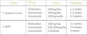
RECOMMENDED DOSING OF ORAL ANTIFUNGALS IN DERMATOPHYCOSES:

9


MOLLUSCUM CONTAGIOSUM (MC):

A cutaneous viral infection caused by pox virus, the disease presents as small pink to white umbilicated papules commonly on face, intertriginous areas and genitalia. Till date 4 viral strains have been isolated causing MC. In healthy young adults disease usually spreads by fomite transfer like infected gym equipment, infected floors and sharing of personal items like towels and soaps.
The incubation period is 3-6 weeks during which intra-cytoplasmic viral replication occurs forming inclusion bodies and causing enlargement of infected cells. The virus tends to be limited to the dermis. In 10% of cases molluscum dermatitis develops. The virus evades immunity by production of virus-specific proteins. Antibody production occurs but is not helpful, while CMI is protective.
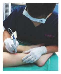
The prognosis is excellent in healthy individuals, with even spontaneous resolution in few. The disease treatment can be done by a variety of methods. Topical treatment includes imquimod cream, cantharidin, tretinoin, TCA, salicylic-lactic acid, silver nitrate, topical cidofovir, rifampicin, podophyllin and potassium hydroxide. Curretage remains the most effective treatment in limited lesions. Cryotherapy and electrocautery can be done too. Intralesional interferon, autoinoculation have been tried in few studies. A sinecatechin Veregen, a green tea extract is being used off-label as 15% paste thrice daily.

VERRUCAE VULGARIS (WARTS):

warts are benign proliferation of the skin and mucosa caused by Human Papilloma Virus (HPV). Till date more than 100 types of HPV have been isolated. Virus tends to infect upper layers of epidermis and cause proliferation. Warts can plane, filirorm, anogenital, mucosal and palmoplantar. Sportsperson and athletes most frequently tend to develop palmoplantar and plane warts from sharing personal items and infected gym equipment. Disease spreads via fomite transfer, autoinoculation and virus enters by superficial abrasions. Incubation period is long usually 3-9 months and relapses and recurrences are frequent.HPV 16, 18, 9, 11, 30, 35 have oncogenic potential.
Common warts present as hyperkeratotic papules with irregular surface and varying sizes. Palmoplantar warts, also called as myrmecia are usually deep well-defined keratotic lesions with a callused periphery. They tend to be deep, painful, on weight bearing areas and resistant to treatment.

TREATMENT –

Topical agents used are 70% salicylic acid, salicylic-lactic acid combination, 5- fluorouracil, TCA, imiquimod, tretinoin, cantharidin, podophylln, contact sensitisers like diphencycloprenone and squaric acid dibutyl ester, aminolevulinic acid with blue light and cidofovir.
Intralesional bleomycin, interferon, candidial antigen, MMR or BCG vaccine have also shown success.
Resistant cases especially palmoplantar can be treated with liquid nitrogen cryotherapy, electrocautery, surgical paring and removal.
Veregen, a green tea sinecatechin extract as 15% paste thrice daily has been FDA approved for treatment of warts.
PDA with ALA and CO2 laser have been tried but their use and safety is debatable.

PIEZOGENIC PEDAL PAPULES:

Resulting due to herniation of fat via fascia into the dermis, these present as small tender firm and compressible papules on the sides of feet and occasionally wrist. Treatment includes wearing orthotic shoes, weight loss, avoidance of prolonged standing and analgesics but permanent relief is usually not achieved. Intralesional infiltration of bupivacaine with betamethasone has shown promise.

CALLOSITY AND CORN: Callus is a hyperkeratotic plaque on

Callus is a hyperkeratotic plaque on site of repetitive pressure, trauma or friction. It is one of the most common sport specific dermatoses. Corn is localised hyperkeratotic tender lesion with a central core on bony prominences. Treatment involves use of keratolytics, paring, pressure relieving orthotic shoes, radiofrequency removal and rarely surgical correction. Corns and calluses can be a very stubborn and physically handicapping disability and should be treated in conjunction with an orthopedic specialist and physiotherapist.

TALON NOIR:

Also called as black heel, it is caused by intra-epidermal haemorrhage over heel and calluses secondary to microtrauma of vasculature secondary to lateral shearing forces. It presents as black dyspigmentation over the affected area. Treatment includes reassurance, analgesics and paring, if required.

JOGGERS’S TOE (tennis toe):

Seen as subungual haematoma in treadmill runners precipitated by rapid starting or stopping, it needs to be differentiated from acral lentiginous melanoma. Reassurance is the key.

FRICTION BLISTER:

These occur due to repetitive shearing forces on opposing surfaces especially with new shoes or extensive use of dumbbells and weights. Presenting as fluid filled blisters of various sizes on both feet or hands, they can be prevented by using cushioned gloves, well fitting shoes and socks. Blisters can be punctured using sterile needle and topical antibiotic with rest of affected area for a couple of days help for healing.

STRIAE DISTENSAE:

Striae occur due to extensive stretching of elastic fibres thanks to sudden weight fluctuation or anabolic steroid usage and are commonly seen on back, arms and legs. Striae are extremely resistant to treatment. TCA application, topical tretinoin, microneedling, PRP and fractional CO2 and erbium-glass laser have been tried with varying rates of success.

SWEAT DERMATITIS:

This form of dermatitis was originally reported from western Rajasthan and is usually ignored or misdiagnosed. Associated with profuse sweating in summers, this disease presents with discolouration and roughening of skin to give a ‘charred appearance’ on trunk, neck and axillary folds. No obstruction of sweat glands is seen. Treatment includes application of mild topical corticosteroid lotions for a few days with oral antihistaminics[3].

URTICARIA AND ANAPHYLAXIS:

Cholinergic urticaria in the form of monomorphic itchy papules can occur due to workout induced sweating. Heat induced urticaria and angioedema can also rarely be precipitated by exercise. Exercise induced anaphylxis can occur in the susceptible especially if workout is done within an hour of food ingestion. Treatment includes antihistaminics, short course corticosteroids and leukotriene antagonists.

INTERTRIGO:

It is an inflammatory dermatoses of skin folds aggravated by friction, moisture, sweating and occlusion by tight clothes. Superinfection by candida most commonly, or bacteria can worsen the clinical picture. Clinically erythematous weeping lesions with satellite lesions and severe burning and itching is seen in crural folds, axilla and infra-mammary area. Lesionscan be relapsing and recurring. Irritant/ allergic dermatitis to synthetic gym wear and arm/ knee bands can also lead to eczematous lesions
Treatment needs to be tailored based on individual causes and keeping affected area dry .is the key. Inciting factors need to be avoided.

CONCLUSION:

Gym is a great avenue for not only staying fit but a social meeting place too. Given its communal nature, it plays host to a multitude of dermatoses, infectious and non-infectious. A dermatologist must keep his mind and eyes open for any clues to the above so that timely intervention can be done.

BASIC PREVENTIVE MEASURES
1.Take bath as soon as possible post workout.
2.Use of liquid handwashes and sanitisers during workout should be encouraged.
3. Use of well fitted and cushioned shoes with change of cotton absorbable socks daily.
4. Avoid sharing of personal products like towels.
5. Disinfect machine handles, weights and dumbbells regularly and use of cushioned cloves can be stressed upon.
6. Use of cotton gym wear.

SUGGESTED READINGS:
1. Nagaraju U, Bhat G, Kuruvila M, Pai GS, Jayalakshmi, Babu RP. Methicillin-resistant Staphylococcus aureus in community-acquired pyoderma. Int J Dermatol. 2004 Jun;43(6):412-4.
2. Venniyil PV, Ganguly S, Kuruvila S, Devi S. A study of community-associated methicillin-resistantStaphylococcus aureus in patients with pyoderma. Indian Dermatol Online J 2016;7:159-63

3. Mehta RD, Bumb RA. Sweat dermatitis. Int J Dermatol.2000 Nov;39(11):872.


The A- Z of HIFU

In today’s fast paced life, patients have been more inclined towards skin tightening and rejuvenation procedures with little or no downtime. The current trend is to opt for noninvasive procedures which offer superior and long lasting results comparable to surgical treatments but without the risk or downtime associated with surgery.There are several treatmentoptions available ranging from non-invasive, minimally – invasive to surgical options. Earlier treatment options ranged from light based or energy based devices like lasers & Radiofrequency which had their own limitations as they delivered heat more superficially. These procedures provide a variable amount of skin tightening but most of them require multiple sessions.

With the advent of High Intensity Focused Ultrasound (HIFU) we are now able to treat deeper layers of the skin in a way that only surgery could treat before. HIFU provides an instant uplift to the skin & more longlasting results.

What is HIFU

HIFU is currently the only non-invasive medical device that uses focused ultrasound energy to selectively heat the superficial and deep dermis as well as the SMAS layer. The SMAS is a subcutaneous, fan – shaped structure that covers the face & connects the facial muscles with the dermis. It has a more durable holding property & less delayed relaxation after lifting procedures than the skin alone thus making it a desirable target for noninvasive skin tightening procedures.HIFU utilises ultrasound energy which is an oscillating pressure wave and can be focused like a beam with a high degree of precision. It is able to reach a deeper depth than the other devices.

Mechanism of Action

The ultrasound waves penetrate into tissue, leading to vibration in molecules at the site of beam focus. The friction between molecules produce heat and thermal injury at the focal site of the beam. Penetration depth is determined by frequency in which higher frequency waves produce a shallow focal injury zone or point and lower frequency waves have a greater depth of penetration to produce focal thermal injury zones (TIZ’s) at deeper layers.At frequency of 1-7 MHZ it can increase the temperature to more than 60 degrees C within the focal zone causing tissue proteins to coagulate followed by neocollagenesis which results in tightening & lifting of the skin. An important aspect of HIFU is that the tissue above & below the focal zone will remain unaffected.

HIFU System Variables

1.Frequency

Higher the frequency more is the absorption of energy & lesser the penetration. Lower the frequency lower is absorption in the tissue and deeper the penetration.

7 Mhz frequency is more superficial & is used for thinner tissue

(e.g. forehead and periorbital areas).

It reaches a depth of 1.5 & 3 mm. It targets the superficial and deep dermis.

4 Mhz frequency reaches deeper layers and is used for thicker tissue (e.g.cheeks & jawline).

It reaches a depth of 4.5 mm.It targets the SMAS layer.

2.Pitch

Pitch implies the distance between the 2 focal thermal coagulative zones or points. When you decrease the pitch the treatment becomes more intense or aggressive. If the spacing between 2 focal points is narrow the chances of side effects could also increase.

3.Energy

Energy indicates the intensity of HIFU. The energy is delivered in a straight line of 5 to 30 mm length. The energy needs to be adjusted according to the thickness of the skin & area to be treated.

Indications:

  • Forehead lines
  • Crows feet
  • Lifting of eyebrows
  • Jowls
  • Neck lines
  • Double Chin
  • Facial Contouring

Contraindications:

  • Pregnancy
  • Bleeding disorders
  • Autoimmune disease
  • Epilepsy
  • Metal implants in the area to be treated
  • Active infection at the sight of treatment

Procedure 

  • Cleanse the skin
  • Take photographs & consent before the treatment
  • Apply topical anaesthesia 30-40 mins prior to the treatment (optional)
  • Mark the preoperative treatment areas with a marker pencil if required.
  • According to the skin thickness select the appropriated cartridge.
  • Set suitable intensity (energy),focal length and pitch.
  • Apply a generous amount of gel on the preoperative area.
  • Maintain complete contact of the cartridge on the skin at all times and do not lift the hand piece during the procedure while the shots are being delivered.
  • Avoid treatment over the temporal branch of trigeminal nerve, marginal mandibular nerve and centre of neck ( thyroid gland).
  • Post procedure sight redness or swelling might be visible which usually subsides within a few hours. Apply moisturizer and sunscreen post procedure.
  • Treatment lines of the ultrasound pulses should be adjacent and parallel to one another with minimal spacing. Vertical and horizontal passes are given in the areas concerned. The total number of lines depends upon size of the treatment area. Approximately 600-800 lines are required for the full face.
  • 5 mm cartridge can be used for areas like cheeks and jawline.This can be followed by a 2nd pass with 3mm cartridge.
  • The 3mm superficial cartridge can be used for areas like neck, double chin and lower face.
  • Additional pass with 1.5mm could be given if required in clients who have fine lines and wrinkles. For eg. Periorbital area, eyebrow and forehead.

Prevention of complications

HIFU

Post treatment erythema is a common occurrence. It usually subsides within the first few hours.

Pain & tenderness is usually experienced around the bony areas like the jawline & the forehead. Application of numbing cream & anxiolytics or pain killers could be opted for in case of clients with a low pain threshold.

Nerve injury can be avoided by not treating the areas where the course of the  nerves becomes relatively superficial.

Geometric wheals or striations generally occur with superficial transducer. Apply ice & topical steroid cream post treatment for fast resolution.

Results

The results are visible immediately as a result of tissue contraction which keep on gradually improving over the next few months. Maximum results are appreciated after 6 to 8 weeks.

The best part about the technology is that it does not require multiple sessions. Depending on the age group & the indication one may require 1 to 2 sessions only.

HIFU has become the most sought after treatment as it offers a significant instant uplift to the skin with no downtime & long lasting results. As it is a safe, effective & non-invasive procedure which offers superior quality of result it also ensures high patient satisfaction range.